
- 阻害剤
- 研究分野別
- PI3K/Akt/mTOR
- Epigenetics
- Methylation
- Immunology & Inflammation
- Protein Tyrosine Kinase
- Angiogenesis
- Apoptosis
- Autophagy
- ER stress & UPR
- JAK/STAT
- MAPK
- Cytoskeletal Signaling
- Cell Cycle
- TGF-beta/Smad
- 化合物ライブラリー
- 抗体
- 新製品
- お問い合わせ
Phospho-LKB1 (Ser428) Antibody [H12K23]
Catalog No.: F0399
Application:
Reactivity:
-
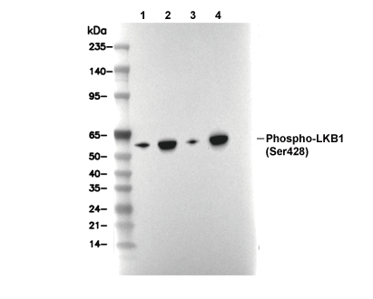 Lane 1: L929, Lane 2: L929 (TPA and UV treated), Lane 3: COS-7, Lane 4: COS-7 (TPA and UV treated)
当該製品は品切れ状态で、メールアドレスをご教示いただければ、お客様に返信いたします。
代表番号: 045-509-1970|電子メール:sales@selleck.co.jp
使用情報
| Dilution |
|---|
|
| Application |
|---|
| WB |
| Source |
|---|
| Rabbit Monoclonal Antibody |
| Reactivity |
|---|
| Human, Mouse, Rat, Monkey |
| Storage Buffer |
|---|
| PBS, pH 7.2+50% Glycerol+0.05% BSA+0.01% NaN3 |
| Storage (from the date of receipt) |
|---|
| -20°C (avoid freeze-thaw cycles), 2 years |
| Predicted MW |
|---|
| 54 kDa |
| ポジティブコントロール | Caki cells (TPA and UV treated); L929 cells (TPA and UV treated); COS cells (TPA and UV treated); NBT-II cells (TPA and UV treated) |
|---|---|
| ネガティブコントロール | Caki cells; NBT-II cells |
プロトコール
| WB |
|---|
Experimental Protocol:
Sample preparation
1. Tissue: Lyse the tissue sample by adding an appropriate volume of ice-cold RIPA/NP-40 Lysis Buffer (containing Protease Inhibitor Cocktail, Phosphatase Inhibitor Cocktail),and homogenize the tissue at a low temperature or lyse it by sonication on ice, then incubate on ice for 30 minutes. 2. Adherent cell: Aspirate the culture medium and wash the cells with ice-cold PBS twice. Lyse the cells by adding an appropriate volume of RIPA/NP-40 Lysis Buffer (containing Protease Inhibitor Cocktail, Phosphatase Inhibitor Cocktail), sonicate to lyse the cells, and incubate on ice for 30 minutes. 3. Suspension cell: Transfer the culture medium to a pre-cooled centrifuge tube. Centrifuge and aspirate the supernatant. Wash the cells with ice-cold PBS twice. Lyse the cells by adding an appropriate volume of RIPA/NP-40 Lysis Buffer (containing Protease Inhibitor Cocktail, Phosphatase Inhibitor Cocktail), sonicate to lyse the cells, and incubate on ice for 30 minutes. 4. Place the lysate into a pre-cooled microcentrifuge tube. Centrifuge at 4°C for 15 min. Collect the supernatant;
5. Remove a small volume of lysate to determine the protein concentration;
6. Combine the lysate with protein loading buffer. Boil 20 µL sample under 95-100°C for 5 min. Centrifuge for 5 min after cool down on ice.
Electrophoretic separation
1. According to the concentration of extracted protein, load appropriate amount of protein sample and marker onto SDS-PAGE gels for electrophoresis. Recommended separating gel (lower gel) concentration: 10%. Reference Table for Selecting SDS-PAGE Separation Gel Concentrations 2. Power up 80V for 30 minutes. Then the power supply is adjusted (110 V~150 V), the Marker is observed, and the electrophoresis can be stopped when the indicator band of the predyed protein Marker where the protein is located is properly separated. (Note that the current should not be too large when electrophoresis, too large current (more than 150 mA) will cause the temperature to rise, affecting the result of running glue. If high currents cannot be avoided, an ice bath can be used to cool the bath.)
Transfer membrane
1. Take out the converter, soak the clip and consumables in the pre-cooled converter;
2. Activate PVDF membrane with methanol for 1 min and rinse with transfer buffer;
3. Install it in the order of "black edge of clip - sponge - filter paper - filter paper - glue -PVDF membrane - filter paper - filter paper - sponge - white edge of clip"; 4. The protein was electrotransferred to PVDF membrane. ( 0.45 µm PVDF membrane is recommended ) Reference Table for Selecting PVDF Membrane Pore Size Specifications Recommended conditions for wet transfer: 200 mA, 120 min. ( Note that the transfer conditions can be adjusted according to the protein size. For high-molecular-weight proteins, a higher current and longer transfer time are recommended. However, ensure that the transfer tank remains at a low temperature to prevent gel melting.)
Block
1. After electrotransfer, wash the film with TBST at room temperature for 5 minutes;
2. Incubate the film in the blocking solution ( recommending 5% BSA solution)
for 1 hour at room temperature;
3. Wash the film with TBST for 3 times, 5 minutes each time.
Antibody incubation
1. Use 5% skim milk powder to prepare the primary antibody working liquid (recommended dilution ratio for primary antibody 1:1000), gently shake and incubate with the film at 4°C overnight; 2. Wash the film with TBST 3 times, 5 minutes each time;
3. Add the secondary antibody to the blocking solution and incubate with the film gently at room temperature for 1 hour;
4. After incubation, wash the film with TBST 3 times for 5 minutes each time.
Antibody staining
1. Add the prepared ECL luminescent substrate (or select other color developing substrate according to the second antibody) and mix evenly;
2. Incubate with the film for 1 minute, remove excess substrate (keep the film moist), wrap with plastic film, and expose in the imaging system. |
生物学的記述
| Specificity |
|---|
| Phospho-LKB1 (Ser428) Antibody [H12K23] detects endogenous levels of total LKB1 protein only when it is phosphorylated at Ser428. |
| タンパク質の局在 |
|---|
| 細胞質、細胞内膜系、ミトコンドリア、細胞核 |
| Uniprot ID |
|---|
| Q15831 |
| Clone |
|---|
| H12K23 |
| Synonym(s) |
|---|
| STK11; Serine/threonine-protein kinase STK11; EC:2.7.11.1; Liver kinase B1 (LKB1; hLKB1); Renal carcinoma antigen NY-REN-19; LKB1; PJS. |
| Background |
|---|
| Phospho-LKB1 (Ser428) represents the activated form of the serine/threonine tumor suppressor kinase LKB1 (STK11), which forms a heterotrimeric complex with STRADα/β and MO25 to phosphorylate and activate AMPK at Thr172 and related kinases, maintaining cellular polarity, energy homeostasis, and growth control. LKB1 contains an N-terminal nuclear localization domain, a central kinase domain with a conserved ATP-binding lysine and activation loop motifs, and a C-terminal non-kinase region that includes Ser428 within a PKCζ consensus site alongside regulatory residues such as Ser325 and Thr363. Flexible loops facilitate the binding of pseudokinase STRAD, enabling allosteric activation. Phosphorylation of Ser428 by PKCζ, in response to signals like metformin treatment, oxidative stress, or energy depletion, triggers export of LKB1 from the nucleus to the cytosol, enhances the stability of the STRAD/MO25 complex, increases catalytic activity, and promotes robust AMPK Thr172 phosphorylation, thereby driving catabolic metabolism, autophagy, and inhibition of mTORC1 and GSK3β. This modification relieves nuclear retention, facilitates LKB1-AMPK docking, suppresses Akt signaling via PTEN stabilization, and fine-tunes apoptotic responses under nutrient stress. Mutation of Ser428 to alanine blocks LKB1 translocation, abolishes metformin-enhanced AMPK activation, and impairs downstream polarity signaling via MARKs and Sad kinases. Dysregulation of LKB1 Ser428 phosphorylation is implicated in diseases such as Peutz-Jeghers syndrome and various cancers, including lung cancer and melanoma, by disrupting energy sensing and growth control, while hyperactivation is associated with endothelial dysfunction in diabetes, and hypoactivation promotes tumorigenesis through unchecked cell proliferation. |
| References |
|---|
技術サポート
ストックの作り方、阻害剤の保管方法、細胞実験や動物実験の際に注意すべき点など、製品を取扱う時に問い合わせが多かった質問に対しては取扱説明書でお答えしています。
他に質問がある場合は、お気軽にお問い合わせください。
* 必須
納期 国内在庫品:受注日の翌日(15時までの受注分) *北海道、九州、沖縄への配送は受注日より2日以上 を要する場合あり 海外在庫品:受注後1〜2週間