
- 阻害剤
- 研究分野別
- PI3K/Akt/mTOR
- Epigenetics
- Methylation
- Immunology & Inflammation
- Protein Tyrosine Kinase
- Angiogenesis
- Apoptosis
- Autophagy
- ER stress & UPR
- JAK/STAT
- MAPK
- Cytoskeletal Signaling
- Cell Cycle
- TGF-beta/Smad
- 化合物ライブラリー
- 抗体
- 新製品
- お問い合わせ
Sec24D Antibody [P23J18]
Catalog No.: F4244
Application:
Reactivity:
-
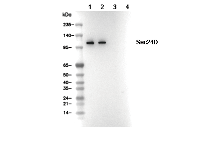 Lane 1: MCF7, Lane 2: Hela, Lane 3: SK-OV-3, Lane 4: 3T3
キーポイント
WB
SDS-PAGE の分離ゲルの推奨濃度:5%。
SDS-PAGE の分離ゲルの推奨濃度:5%。
使用情報
| Dilution |
|---|
|
| Application |
|---|
| WB, IP |
| Source |
|---|
| Rabbit Monoclonal Antibody |
| Reactivity |
|---|
| Human, Mouse, Rat, Monkey |
| Storage Buffer |
|---|
| PBS, pH 7.2+50% Glycerol+0.05% BSA+0.01% NaN3 |
| Storage (from the date of receipt) |
|---|
| -20°C (avoid freeze-thaw cycles), 2 years |
| Predicted MW |
|---|
| 115 kDa |
| ポジティブコントロール | HeLa cell; MCF-7 cell; SK-OV-3 cell; COS-7 cell; 3T3 cell; PC12 cell |
|---|---|
| ネガティブコントロール |
プロトコール
| WB |
|---|
Experimental Protocol:
Sample preparation
1. Tissue: Lyse the tissue sample by adding an appropriate volume of ice-cold RIPA/NP-40 Lysis Buffer (containing Protease Inhibitor Cocktail),and homogenize the tissue at a low temperature or lyse it by sonication on ice, then incubate on ice for 30 minutes. 2. Adherent cell: Aspirate the culture medium and wash the cells with ice-cold PBS twice. Lyse the cells by adding an appropriate volume of RIPA/NP-40 Lysis Buffer (containing Protease Inhibitor Cocktail), sonicate to lyse the cells, and incubate on ice for 30 minutes. 3. Suspension cell: Transfer the culture medium to a pre-cooled centrifuge tube. Centrifuge and aspirate the supernatant. Wash the cells with ice-cold PBS twice. Lyse the cells by adding an appropriate volume of RIPA/NP-40 Lysis Buffer (containing Protease Inhibitor Cocktail), sonicate to lyse the cells, and incubate on ice for 30 minutes. 4. Place the lysate into a pre-cooled microcentrifuge tube. Centrifuge at 4°C for 15 min. Collect the supernatant;
5. Remove a small volume of lysate to determine the protein concentration;
6. Combine the lysate with protein loading buffer. Boil 20 µL sample under 95-100°C for 5 min. Centrifuge for 5 min after cool down on ice.
Electrophoretic separation
1. According to the concentration of extracted protein, load appropriate amount of protein sample and marker onto SDS-PAGE gels for electrophoresis. Recommended separating gel (lower gel) concentration: 5%. Reference Table for Selecting SDS-PAGE Separation Gel Concentrations 2. Power up 80V for 30 minutes. Then the power supply is adjusted (110 V~150 V), the Marker is observed, and the electrophoresis can be stopped when the indicator band of the predyed protein Marker where the protein is located is properly separated. (Note that the current should not be too large when electrophoresis, too large current (more than 150 mA) will cause the temperature to rise, affecting the result of running glue. If high currents cannot be avoided, an ice bath can be used to cool the bath.)
Transfer membrane
1. Take out the converter, soak the clip and consumables in the pre-cooled converter;
2. Activate PVDF membrane with methanol for 1 min and rinse with transfer buffer;
3. Install it in the order of "black edge of clip - sponge - filter paper - filter paper - glue -PVDF membrane - filter paper - filter paper - sponge - white edge of clip"; 4. The protein was electrotransferred to PVDF membrane. ( 0.45 µm PVDF membrane is recommended ) Reference Table for Selecting PVDF Membrane Pore Size Specifications Recommended conditions for wet transfer: 200 mA, 120 min. ( Note that the transfer conditions can be adjusted according to the protein size. For high-molecular-weight proteins, a higher current and longer transfer time are recommended. However, ensure that the transfer tank remains at a low temperature to prevent gel melting.)
Block
1. After electrotransfer, wash the film with TBST at room temperature for 5 minutes;
2. Incubate the film in the blocking solution for 1 hour at room temperature;
3. Wash the film with TBST for 3 times, 5 minutes each time.
Antibody incubation
1. Use 5% skim milk powder to prepare the primary antibody working liquid (recommended dilution ratio for primary antibody 1:1000), gently shake and incubate with the film at 4°C overnight; 2. Wash the film with TBST 3 times, 5 minutes each time;
3. Add the secondary antibody to the blocking solution and incubate with the film gently at room temperature for 1 hour;
4. After incubation, wash the film with TBST 3 times for 5 minutes each time.
Antibody staining
1. Add the prepared ECL luminescent substrate (or select other color developing substrate according to the second antibody) and mix evenly;
2. Incubate with the film for 1 minute, remove excess substrate (keep the film moist), wrap with plastic film, and expose in the imaging system. |
生物学的記述
| Specificity |
|---|
| Sec24D Antibody [P23J18] detects endogenous levels of total Sec24D protein. |
| タンパク質の局在 |
|---|
| 細胞質、細胞質小胞、小胞体、細胞内膜系 |
| Uniprot ID |
|---|
| O94855 |
| Clone |
|---|
| P23J18 |
| Synonym(s) |
|---|
| Protein transport protein Sec24D; SEC24-related protein D; SEC24D; KIAA0755. |
| Background |
|---|
| SEC24D (SEC24 Homolog D, COPII Coat Complex Component) is a member of the SEC24 gene subfamily and encodes a core structural and functional element of the COPII (Coat Protein Complex II) machinery, which mediates the vesicular transport of newly synthesized proteins from the endoplasmic reticulum (ER) to the Golgi apparatus. The assembly of COPII-coated vesicles is initiated when the small GTPase Sar1 interacts with the Sec23/24 complex, generating a prebudding intermediate that selectively captures cargo destined for secretion. Within this complex, the Sec24 subunit is primarily responsible for cargo recognition and selection, as it directly engages specific export signals on transmembrane or soluble proteins within the ER lumen. These interactions ensure efficient incorporation of cargo into forming COPII vesicles through cooperative binding with Sec23. In humans, four distinct Sec24 isoforms—Sec24A, Sec24B, Sec24C, and Sec24D have been identified, each displaying unique yet overlapping cargo specificities and tissue expression profiles. Among these, SEC24D plays a vital role in maintaining ER-to-Golgi trafficking fidelity, contributing to proper protein sorting, secretion, and cellular homeostasis. SEC24D gene is overexpressed in metastasis across Kidney Renal Clear Cell Carcinoma (KIRC), Lung Squamous Cell Carcinoma (LUSC), and Stomach Adenocarcinoma (STAD) patients. |
| References |
|---|
技術サポート
ストックの作り方、阻害剤の保管方法、細胞実験や動物実験の際に注意すべき点など、製品を取扱う時に問い合わせが多かった質問に対しては取扱説明書でお答えしています。
他に質問がある場合は、お気軽にお問い合わせください。
* 必須
納期 国内在庫品:受注日の翌日(15時までの受注分) *北海道、九州、沖縄への配送は受注日より2日以上 を要する場合あり 海外在庫品:受注後1〜2週間