
- 阻害剤
- 研究分野別
- PI3K/Akt/mTOR
- Epigenetics
- Methylation
- Immunology & Inflammation
- Protein Tyrosine Kinase
- Angiogenesis
- Apoptosis
- Autophagy
- ER stress & UPR
- JAK/STAT
- MAPK
- Cytoskeletal Signaling
- Cell Cycle
- TGF-beta/Smad
- 化合物ライブラリー
- 抗体
- 新製品
- お問い合わせ
SSEA4 Antibody [E20H6]
Catalog No.: F2673
Application:
Reactivity:
-
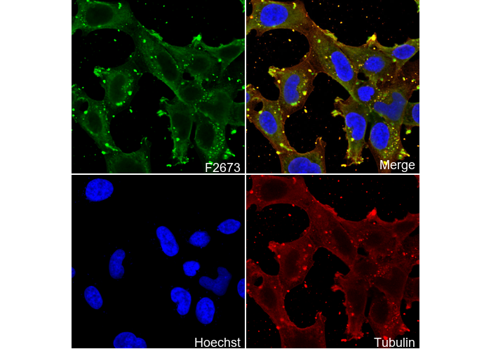 Immunofluorescent analysis of SW480 cells using F2673 (green, 1:100 ), Hoechst (blue) and tubulin (Red).
使用情報
| Dilution |
|---|
|
| Application |
|---|
| IF, FCM |
| Source |
|---|
| Mouse Monoclonal Antibody |
| Reactivity |
|---|
| Human |
| Storage Buffer |
|---|
| PBS, pH 7.2+50% Glycerol+0.05% BSA+0.01% NaN3 |
| Storage (from the date of receipt) |
|---|
| -20°C (avoid freeze-thaw cycles), 2 years |
| ポジティブコントロール | NTERA-2 cells |
|---|---|
| ネガティブコントロール | Jurkat cells |
プロトコール
| IF |
|---|
Experimental Protocol:
Sample Preparation
1. Adherent Cells: Place a clean, sterile coverslip in a culture dish. Once the cells grow to near confluence as a monolayer, remove the coverslip for further use.
2. Suspension Cells: Seed the cells onto a clean, sterile slide coated with poly-L-lysine.
3. Frozen Sections: Allow the slide to thaw at room temperature. Wash it with pure water or PBS for 2 times, 3 minutes each time.
4. Paraffin Sections: Deparaffinization and rehydration. Wash the slide with pure water or PBS for 3 times, 3 minutes each time. Then perform antigen retrieval.
Fixation
1. Fix the cell coverslips/spots or tissue sections at room temperature using a fixative such as 4% paraformaldehyde (4% PFA) for 10-15 minutes.
2. Wash the sample with PBS for 3 times, 3 minutes each time.
Permeabilization
1.Add a detergent such as 0.1–0.3% Triton X-100 to the sample and incubate at room temperature for 10–20 minutes.
(Note: This step is only required for intracellular antigens. For antigens expressed on the cell membrane, this step is unnecessary.)
Wash the sample with PBS for 3 times, 3 minutes each time.
Blocking
Add blocking solution and incubate at room temperature for at least 1 hour. (Common blocking solutions include: serum from the same source as the secondary antibody, BSA, or goat serum.)
Note: Ensure the sample remains moist during and after the blocking step to prevent drying, which can lead to high background.
Immunofluorescence Staining (Day 1)
1. Remove the blocking solution and add the diluted primary antibody.
2. Incubate the sample in a humidified chamber at 4°C overnight.
Immunofluorescence Staining (Day 2)
1. Remove the primary antibody and wash with PBST for 3 times, 5 minutes each time.
2. Add the diluted fluorescent secondary antibody and incubate in the dark at 4°C for 1–2 hours.
3. Remove the secondary antibody and wash with PBST for 3 times, 5 minutes each time.
4. Add diluted DAPI and incubate at room temperature in the dark for 5–10 minutes.
5. Wash with PBST for 3 times, 5 minutes each time.
Mounting
1. Mount the sample with an anti-fade mounting medium.
2. Allow the slide to dry at room temperature overnight in the dark.
3. Store the slide in a slide storage box at 4°C, protected from light.
|
生物学的記述
| Specificity |
|---|
| SSEA4 Antibody [E20H6] detects endogenous levels of total SSEA4 protein. |
| Clone |
|---|
| E20H6 |
| Background |
|---|
| SSEA4 (Stage-Specific Embryonic Antigen 4) is a sialylated hexasaccharide glycolipid epitope (Neu5Acα2-3Galβ1-3GalNAcβ1-3Galα1-4Galβ1-4Glc-Cer) found on human embryonic stem cells (hESCs), induced pluripotent stem cells (iPSCs), teratocarcinoma cells, and cancer stem cells, but absent from differentiated somatic or mouse pluripotent cells. Its carbohydrate headgroup adopts a horseshoe-shaped conformation recognized by MC813 antibodies through a network of hydrogen bonds involving the terminal sialic acid (Neu5Ac) carboxylate with Asp54H/Gly53H and N-acetyl GalNAc hydroxyls with Asp49L, which underpins its utility for specific detection of pluripotency states via flow cytometry and immunohistochemistry. The glycan’s rigid α-Gal linkage and β1-3 glycosidic bonds maintain a stable, extended conformation atop ceramide lipid tails in plasma membranes, where SSEA4 actively supports stemness by modulating Wnt/β-catenin signaling through direct interaction with the LRP6 co-receptor, enabling ligand-independent pathway activation, and by recruiting PI3K/Akt through glycan-mediated clustering that sustains Nanog/Oct4/Sox2 circuitry essential for self-renewal. During differentiation, downregulation of glycosyltransferases (ST3GAL6, B3GALT5) progressively removes the terminal sialic acid, converting SSEA4 to SSEA3 and extinguishing its signaling competence. |
| References |
|---|
技術サポート
ストックの作り方、阻害剤の保管方法、細胞実験や動物実験の際に注意すべき点など、製品を取扱う時に問い合わせが多かった質問に対しては取扱説明書でお答えしています。
他に質問がある場合は、お気軽にお問い合わせください。
* 必須
納期 国内在庫品:受注日の翌日(15時までの受注分) *北海道、九州、沖縄への配送は受注日より2日以上 を要する場合あり 海外在庫品:受注後1〜2週間