
- 阻害剤
- 研究分野別
- PI3K/Akt/mTOR
- Epigenetics
- Methylation
- Immunology & Inflammation
- Protein Tyrosine Kinase
- Angiogenesis
- Apoptosis
- Autophagy
- ER stress & UPR
- JAK/STAT
- MAPK
- Cytoskeletal Signaling
- Cell Cycle
- TGF-beta/Smad
- 化合物ライブラリー
- 抗体
- 新製品
- お問い合わせ
Ubc12 Antibody [K5G17]
Catalog No.: F5062
Application:
Reactivity:
-
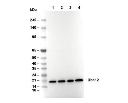 Lane 1: Hela, Lane 2: NIH/3T3, Lane 3: C6, Lane 4: COS-7
当該製品は品切れ状态で、メールアドレスをご教示いただければ、お客様に返信いたします。
代表番号: 045-509-1970|電子メール:sales@selleck.co.jp
キーポイント
WB
転写条件(ウェット): 200 mA, 60 min。
転写条件(ウェット): 200 mA, 60 min。
使用情報
| Dilution |
|---|
|
| Application |
|---|
| WB, IP |
| Source |
|---|
| Rabbit Monoclonal Antibody |
| Reactivity |
|---|
| Human, Mouse, Rat, Monkey |
| Storage Buffer |
|---|
| PBS, pH 7.2+50% Glycerol+0.05% BSA+0.01% NaN3 |
| Storage (from the date of receipt) |
|---|
| -20°C (avoid freeze-thaw cycles), 2 years |
| Predicted MW |
|---|
| 21 kDa |
| ポジティブコントロール | HeLa cells; NIH/3T3 cells; C6 cells; COS-7 cells; 293 cells |
|---|---|
| ネガティブコントロール |
プロトコール
| WB |
|---|
Experimental Protocol:
Sample preparation
1. Tissue: Lyse the tissue sample by adding an appropriate volume of ice-cold RIPA/NP-40 Lysis Buffer (containing Protease Inhibitor Cocktail),and homogenize the tissue at a low temperature. 2. Adherent cell: Aspirate the culture medium and wash the cells with ice-cold PBS twice. Lyse the cells by adding an appropriate volume of RIPA/NP-40 Lysis Buffer (containing Protease Inhibitor Cocktail) and put the sample on ice for 5 min. 3. Suspension cell: Transfer the culture medium to a pre-cooled centrifuge tube. Centrifuge and aspirate the supernatant. Wash the cells with ice-cold PBS twice. Lyse the cells by adding an appropriate volume of RIPA/NP-40 Lysis Buffer (containing Protease Inhibitor Cocktail) and put the sample on ice for 5 min. 4. Place the lysate into a pre-cooled microcentrifuge tube. Centrifuge at 4°C for 15 min. Collect the supernatant;
5. Remove a small volume of lysate to determine the protein concentration;
6. Combine the lysate with protein loading buffer. Boil 20 µL sample under 95-100°C for 5 min. Centrifuge for 5 min after cool down on ice.
Electrophoretic separation
1. According to the concentration of extracted protein, load appropriate amount of protein sample and marker onto SDS-PAGE gels for electrophoresis. Recommended separating gel (lower gel) concentration: 10%. Reference Table for Selecting SDS-PAGE Separation Gel Concentrations 2. Power up 80V for 30 minutes. Then the power supply is adjusted (110 V~150 V), the Marker is observed, and the electrophoresis can be stopped when the indicator band of the predyed protein Marker where the protein is located is properly separated. (Note that the current should not be too large when electrophoresis, too large current (more than 150 mA) will cause the temperature to rise, affecting the result of running glue. If high currents cannot be avoided, an ice bath can be used to cool the bath.)
Transfer membrane
1. Take out the converter, soak the clip and consumables in the pre-cooled converter;
2. Activate PVDF membrane with methanol for 1 min and rinse with transfer buffer;
3. Install it in the order of "black edge of clip - sponge - filter paper - filter paper - glue -PVDF membrane - filter paper - filter paper - sponge - white edge of clip"; 4. The protein was electrotransferred to PVDF membrane. ( 0.45 µm PVDF membrane is recommended ) Reference Table for Selecting PVDF Membrane Pore Size Specifications Recommended conditions for wet transfer: 200 mA, 60 min. ( Note that the transfer conditions can be adjusted according to the protein size. For high-molecular-weight proteins, a higher current and longer transfer time are recommended. However, ensure that the transfer tank remains at a low temperature to prevent gel melting.)
Block
1. After electrotransfer, wash the film with TBST at room temperature for 5 minutes;
2. Incubate the film in the blocking solution for 1 hour at room temperature;
3. Wash the film with TBST for 3 times, 5 minutes each time.
Antibody incubation
1. Use 5% skim milk powder to prepare the primary antibody working liquid (recommended dilution ratio for primary antibody 1:1000), gently shake and incubate with the film at 4°C overnight; 2. Wash the film with TBST 3 times, 5 minutes each time;
3. Add the secondary antibody to the blocking solution and incubate with the film gently at room temperature for 1 hour;
4. After incubation, wash the film with TBST 3 times for 5 minutes each time.
Antibody staining
1. Add the prepared ECL luminescent substrate (or select other color developing substrate according to the second antibody) and mix evenly;
2. Incubate with the film for 1 minute, remove excess substrate (keep the film moist), wrap with plastic film, and expose in the imaging system. |
生物学的記述
| Specificity |
|---|
| Ubc12 Antibody [K5G17] detects endogenous levels of total Ubc12 protein. |
| Uniprot ID |
|---|
| P61081 |
| Clone |
|---|
| K5G17 |
| Synonym(s) |
|---|
| NEDD8-conjugating enzyme Ubc12; NEDD8 carrier protein; Ubiquitin-conjugating enzyme E2 M; UBE2M; UBC12 |
| Background |
|---|
| Ubc12, the dedicated E2-conjugating enzyme for NEDD8 in the neddylation cascade, parallels ubiquitin-conjugating enzymes but specifically activates cullin-RING ligases (CRLs) by transferring NEDD8 to lysine residues on cullin scaffolds, thereby enhancing ubiquitin ligase activity essential for proteostasis. It adopts a canonical UBC fold with a flexible C-terminal extension that facilitates heterodimer formation with NEDD8~E1 thioester, enabling direct transfer to unneddylated cullins following displacement of inhibitory factors like CAND1. Ubc12 charged with NEDD8 promotes cullin conformational remodeling, repositioning the Cul-Rbx1 RING subdomain to recruit ubiquitin E2s such as Cdc34 for processive K48/K63 chain assembly on substrates; in SCF^βTrCP complexes, this neddylation is strictly required for optimal phosphorylation-dependent IκBα recognition, ubiquitination, and proteasomal degradation, unleashing NF-κB p65/p50 nuclear translocation to transcribe proinflammatory cytokines like TNF-α and IL-6, while in SCF^Skp2, it drives p27^Kip1 turnover by stabilizing Skp1-Skp2 docking, licensing CDK2 activation for G1/S transition. Dynamic cycles of neddylation by Ubc12 oppose CSN-mediated deneddylation, with Skp2-Skp1 actively dissociating CAND1 from Cul1 to favor neddylation, whereas bound substrates shield neddylated Cul1 from CSN, sustaining CRL activity during signaling flux. Ubc12 governs immune activation, cell cycle fidelity, and developmental patterning by ensuring timely degradation of NF-κB inhibitors and CDK regulators, making it indispensable for researchers studying CRL dependency in proliferation or inflammation where neddylation inhibition phenocopies cullin loss. Its activity maintains epithelial barrier integrity and hematopoietic homeostasis through NF-κB oscillations, with broad expression supporting conditional knockout models for tissue-specific pathway dissection. Dysregulation via neddylation blockade accumulates IκB and p27, blunting innate responses and arresting proliferation, as evidenced in inflammatory bowel disease models and tumorigenesis. |
| References |
|---|
技術サポート
ストックの作り方、阻害剤の保管方法、細胞実験や動物実験の際に注意すべき点など、製品を取扱う時に問い合わせが多かった質問に対しては取扱説明書でお答えしています。
他に質問がある場合は、お気軽にお問い合わせください。
* 必須
納期 国内在庫品:受注日の翌日(15時までの受注分) *北海道、九州、沖縄への配送は受注日より2日以上 を要する場合あり 海外在庫品:受注後1〜2週間