
- 阻害剤
- 研究分野別
- PI3K/Akt/mTOR
- Epigenetics
- Methylation
- Immunology & Inflammation
- Protein Tyrosine Kinase
- Angiogenesis
- Apoptosis
- Autophagy
- ER stress & UPR
- JAK/STAT
- MAPK
- Cytoskeletal Signaling
- Cell Cycle
- TGF-beta/Smad
- 化合物ライブラリー
- 抗体
- 新製品
- お問い合わせ

A1/Bfl-1 Antibody (Rabbit mAb) [N2F22]
CatNo: F5067
Application:
Reactivity:
-
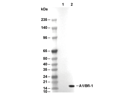 Lane 1: 293T, Lane 2: 293T (BCL2A1(human) transfected)
当該製品は品切れ状态で、メールアドレスをご教示いただければ、お客様に返信いたします。
代表番号: 045-509-1970|電子メール:sales@selleck.co.jp
キーポイント
WB
転写条件(ウェット): 200 mA, 60 min, 0.22 μm PVDF 膜の使用をお勧めします。
転写条件(ウェット): 200 mA, 60 min, 0.22 μm PVDF 膜の使用をお勧めします。
使用情報
| Dilution |
|---|
|
| Application |
|---|
| WB, IP |
| Source |
|---|
| Rabbit Monoclonal Antibody |
| Reactivity |
|---|
| Human |
| Storage Buffer |
|---|
| PBS, pH 7.2+50% Glycerol+0.05% BSA+0.01% NaN3 |
| Storage (from the date of receipt) |
|---|
| -20°C (avoid freeze-thaw cycles), 2 years |
| Predicted MW |
|---|
| 18 kDa |
| ポジティブコントロール | U-937 cells; UACC-62 cells; Mutz-3 cells |
|---|---|
| ネガティブコントロール |
プロトコール
| WB |
|---|
Experimental Protocol:
Sample preparation
1. Tissue: Lyse the tissue sample by adding an appropriate volume of ice-cold RIPA/Tris-HCL Lysis Buffer (containing Protease Inhibitor Cocktail),and homogenize the tissue at a low temperature or lyse it by sonication on ice, then incubate on ice for 30 minutes. 2. Adherent cell: Aspirate the culture medium and wash the cells with ice-cold PBS twice. Lyse the cells by adding an appropriate volume of RIPA/Tris-HCL Lysis Buffer (containing Protease Inhibitor Cocktail) , sonicate to lyse the cells, and incubate on ice for 30 minutes. 3. Suspension cell: Transfer the culture medium to a pre-cooled centrifuge tube. Centrifuge and aspirate the supernatant. Wash the cells with ice-cold PBS twice. Lyse the cells by adding an appropriate volume of RIPA/Tris-HCL Lysis Buffer (containing Protease Inhibitor Cocktail) , sonicate to lyse the cells, and incubate on ice for 30 minutes. 4. Place the lysate into a pre-cooled microcentrifuge tube. Centrifuge at 4°C for 15 min. Collect the supernatant;
5. Remove a small volume of lysate to determine the protein concentration;
6. Combine the lysate with protein loading buffer. Boil 20 µL sample under 95-100°C for 5 min. Centrifuge for 5 min after cool down on ice.
Electrophoretic separation
1. According to the concentration of extracted protein, load appropriate amount of protein sample and marker onto SDS-PAGE gels for electrophoresis. Recommended separating gel (lower gel) concentration: 10%. Reference Table for Selecting SDS-PAGE Separation Gel Concentrations 2. Power up 80V for 30 minutes. Then the power supply is adjusted (110 V~150 V), the Marker is observed, and the electrophoresis can be stopped when the indicator band of the predyed protein Marker where the protein is located is properly separated. (Note that the current should not be too large when electrophoresis, too large current (more than 150 mA) will cause the temperature to rise, affecting the result of running glue. If high currents cannot be avoided, an ice bath can be used to cool the bath.)
Transfer membrane
1. Take out the converter, soak the clip and consumables in the pre-cooled converter;
2. Activate PVDF membrane with methanol for 1 min and rinse with transfer buffer;
3. Install it in the order of "black edge of clip - sponge - filter paper - filter paper - glue -PVDF membrane - filter paper - filter paper - sponge - white edge of clip"; 4. The protein was electrotransferred to PVDF membrane. ( 0.22 µm PVDF membrane is recommended )) Reference Table for Selecting PVDF Membrane Pore Size Specifications Recommended conditions for wet transfer: 200 mA, 60 min. ( Note that the transfer conditions can be adjusted according to the protein size. For high-molecular-weight proteins, a higher current and longer transfer time are recommended. However, ensure that the transfer tank remains at a low temperature to prevent gel melting.)
Block
1. After electrotransfer, wash the film with TBST at room temperature for 5 minutes;
2. Incubate the film in the blocking solution for 1 hour at room temperature;
3. Wash the film with TBST for 3 times, 5 minutes each time.
Antibody incubation
1. Use primary antibody dilution buffer to prepare the primary antibody working liquid (recommended dilution ratio for primary antibody 1:1000), gently shake and incubate with the film at 4°C overnight; 2. Wash the film with TBST 3 times, 5 minutes each time;
3. Add the secondary antibody to the blocking solution and incubate with the film gently at room temperature for 1 hour;
4. After incubation, wash the film with TBST 3 times for 5 minutes each time.
Antibody staining
1. Add the prepared ECL luminescent substrate (or select other color developing substrate according to the second antibody) and mix evenly;
2. Incubate with the film for 1 minute, remove excess substrate (keep the film moist), wrap with plastic film, and expose in the imaging system. |
生物学的記述
| Specificity |
|---|
| A1/Bfl-1 Antibody (Rabbit mAb) [N2F22] detects endogenous levels of total A1/Bfl-1 protein. |
| タンパク質の局在 |
|---|
| 細胞質 |
| Uniprot ID |
|---|
| Q16548 |
| Clone |
|---|
| N2F22 |
| Synonym(s) |
|---|
| ACC-1; ACC-2; ACC1; ACC2; B2LA1; BCL2A1; BCL2L5; BFL1; GRS; HBPA1; hematopoietic BCL2-related protein A1; Hemopoietic-specific early response protein; Protein BFL-1; Protein GRS |
| Background |
|---|
| A1/Bfl-1 (BCL2A1) represents an anti-apoptotic member of the Bcl-2 protein family originally identified as a granulocyte-macrophage colony-stimulating factor (GM-CSF)-inducible gene in mouse bone marrow, exhibiting predominant expression in hematopoietic cells, including neutrophils, macrophages, T lymphocytes, and B lymphocytes, with limited detection in non-hematopoietic tissues such as lung and vascular endothelium. A1/Bfl-1 contains four Bcl-2 homology (BH) domains organized into eight alpha-helices, with helices α4, α5, and α6 corresponding to BH3, BH1, and BH2 domains forming a hydrophobic groove on the protein surface for interaction with pro-apoptotic Bcl-2 family members, although A1 uniquely possesses an acidic glutamate residue at position 78 within this binding groove distinguishing its interaction specificity from other anti-apoptotic relatives. The protein diverges structurally from Bcl-2, Bcl-xL, Bcl-w, and Mcl-1 through its hydrophilic C-terminal domain that lacks a transmembrane anchor, instead containing ubiquitination sites directing rapid proteasomal degradation conferring a short half-life of approximately thirty minutes, comparable to Mcl-1 but contrasting with Bcl-2's twenty-four-hour stability. A1/Bfl-1 expression becomes rapidly induced through NF-κB transcriptional activation in response to diverse inflammatory stimuli, including TNF-α, IL-1β, CD40 ligation, phorbol esters, lipopolysaccharide, and pre-T-cell receptor signaling operating through PLCγ/PKC/NF-κB pathways, positioning A1 as an immediate-early survival response gene. The protein functions by binding and neutralizing BH3-only pro-apoptotic proteins, including Bid, Bim, and Puma with high affinity while exhibiting no detectable binding to Bad, thereby preventing these death activators from triggering Bax/Bak oligomerization and mitochondrial outer membrane permeabilization that would otherwise release cytochrome c and initiate caspase-dependent apoptosis. A1/Bfl-1 displays preferential interaction with Bak over Bax through direct binding that antagonizes Bak-mediated cell death, although conflicting reports describe species-specific differences wherein mouse A1 localizes predominantly to the cytosol requiring interaction partners to impose its site of anti-apoptotic action while human A1/Bfl-1 preferentially targets mitochondria directly. The protein executes critical survival functions during lymphocyte development—A1 expression peaks at the DN3 to DN4 thymocyte transition where pre-TCR signaling-dependent A1 induction proves necessary and sufficient for beta-selection progression, reappears in CD4+ single-positive thymocytes at thirty-fold higher levels than double-positive cells, and becomes strongly upregulated upon T-cell receptor engagement in peripheral T cells along with Noxa to enable survival of high-affinity TCR-bearing antigen-specific clones. A1/Bfl-1 regulates mature follicular B-cell survival downstream of B-cell receptor and PLCγ2 signaling through NF-κB activation, rendering follicular B cells resistant to BCR-induced apoptosis that eliminates transitional B cells during negative selection, with elevated A1 expression documented in systemic lupus erythematosus patients' B cells reflecting dysregulated tolerance mechanisms. The protein mediates neutrophil survival extension during bacterial infection and inflammatory responses by countering spontaneous apoptosis in response to G-CSF, GM-CSF, TNF-α, IFN-γ, IL-8, and lipopolysaccharide stimulation through NF-κB-dependent transcription, with loss of A1-a isoform abolishing LPS-mediated neutrophil survival enhancement although steady-state and inflammatory granulocyte numbers remain normal due to compensatory progenitor activity. A1/Bfl-1 promotes macrophage survival during infection with Mycobacterium bovis BCG, Toxoplasma gondii, and Mycobacterium tuberculosis through NF-κB pathway activation peaking within eight to sixteen hours post-infection, yet paradoxically inhibits autophagosome maturation and acidification in virulent M. tuberculosis-infected macrophages, enabling intracellular bacterial persistence by blocking both autophagy-mediated killing and host cell apoptosis. A1 exhibits functional redundancy with Mcl-1 in mast cell survival, collectively mediating allergic reaction severity by sustaining the survival of tissue-resident mast cells containing histamine-rich inflammatory granules. Calpain proteolytically cleaves A1/Bfl-1 within the N-terminal region, generating pro-apoptotic C-terminal fragments that promote mitochondrial cytochrome c release and apoptosis reversal, representing a potential therapeutic mechanism, although physiological contexts triggering this conversion remain incompletely defined. A1/Bfl-1 overexpression fails to induce lymphoma in transgenic mice, unlike Bcl-2, yet ubiquitination-resistant A1 mutants predispose to lymphomagenesis when co-expressed with dominant-negative p53 and accelerate Myc-induced lymphoma development, while elevated A1 expression in B chronic lymphocytic leukemia, acute myeloid leukemia, and acute promyelocytic leukemia strongly correlates with chemoresistance and poor prognosis. |
| References |
|---|
技術サポート
ストックの作り方、阻害剤の保管方法、細胞実験や動物実験の際に注意すべき点など、製品を取扱う時に問い合わせが多かった質問に対しては取扱説明書でお答えしています。
他に質問がある場合は、お気軽にお問い合わせください。
* 必須

納期 国内在庫品:受注日の翌日(15時までの受注分) *北海道、九州、沖縄への配送は受注日より2日以上 を要する場合あり 海外在庫品:受注後1〜2週間