
- 阻害剤
- 研究分野別
- PI3K/Akt/mTOR
- Epigenetics
- Methylation
- Immunology & Inflammation
- Protein Tyrosine Kinase
- Angiogenesis
- Apoptosis
- Autophagy
- ER stress & UPR
- JAK/STAT
- MAPK
- Cytoskeletal Signaling
- Cell Cycle
- TGF-beta/Smad
- 化合物ライブラリー
- 抗体
- 新製品
- お問い合わせ
Atg5 Antibody (Rabbit mAb) [A3G17]
Catalog No.: F0422
Application:
Reactivity:
-
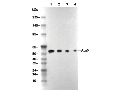 Lane 1: PANC-1, Lane 2: HCT116, Lane 3: H-4-II-E, Lane 4: C2C12
文献中Selleckの製品使用例(1)
使用情報
| Dilution |
|---|
|
| Application |
|---|
| WB, IP |
| Source |
|---|
| Rabbit Monoclonal Antibody |
| Reactivity |
|---|
| Human, Mouse, Rat |
| Storage Buffer |
|---|
| PBS, pH 7.2+50% Glycerol+0.05% BSA+0.01% NaN3 |
| Storage (from the date of receipt) |
|---|
| -20°C (avoid freeze-thaw cycles), 2 years |
| Predicted MW |
|---|
| 50-55 kDa |
| ポジティブコントロール | PANC-1 cells; U-87 MG cells; Saos-2 cells; HCT 116 cells; MCF7 cells; OVCAR8 cells; HeLa cells; C2C12 cells; L-929 cells; C6 cells; H-4-II-E cells; MEFs cells |
|---|---|
| ネガティブコントロール |
プロトコール
| WB |
|---|
Experimental Protocol:
Sample preparation
1. Tissue: Lyse the tissue sample by adding an appropriate volume of ice-cold RIPA/NP-40 Lysis Buffer (containing Protease Inhibitor Cocktail),and homogenize the tissue at a low temperature or lyse it by sonication on ice, then incubate on ice for 30 minutes. 2. Adherent cell: Aspirate the culture medium and wash the cells with ice-cold PBS twice. Lyse the cells by adding an appropriate volume of RIPA/NP-40 Lysis Buffer (containing Protease Inhibitor Cocktail), sonicate to lyse the cells, and incubate on ice for 30 minutes. 3. Suspension cell: Transfer the culture medium to a pre-cooled centrifuge tube. Centrifuge and aspirate the supernatant. Wash the cells with ice-cold PBS twice. Lyse the cells by adding an appropriate volume of RIPA/NP-40 Lysis Buffer (containing Protease Inhibitor Cocktail), sonicate to lyse the cells, and incubate on ice for 30 minutes. 4. Place the lysate into a pre-cooled microcentrifuge tube. Centrifuge at 4°C for 15 min. Collect the supernatant;
5. Remove a small volume of lysate to determine the protein concentration;
6. Combine the lysate with protein loading buffer. Boil 20 µL sample under 95-100°C for 5 min. Centrifuge for 5 min after cool down on ice.
Electrophoretic separation
1. According to the concentration of extracted protein, load appropriate amount of protein sample and marker onto SDS-PAGE gels for electrophoresis. Recommended separating gel (lower gel) concentration: 10%. Reference Table for Selecting SDS-PAGE Separation Gel Concentrations 2. Power up 80V for 30 minutes. Then the power supply is adjusted (110 V~150 V), the Marker is observed, and the electrophoresis can be stopped when the indicator band of the predyed protein Marker where the protein is located is properly separated. (Note that the current should not be too large when electrophoresis, too large current (more than 150 mA) will cause the temperature to rise, affecting the result of running glue. If high currents cannot be avoided, an ice bath can be used to cool the bath.)
Transfer membrane
1. Take out the converter, soak the clip and consumables in the pre-cooled converter;
2. Activate PVDF membrane with methanol for 1 min and rinse with transfer buffer;
3. Install it in the order of "black edge of clip - sponge - filter paper - filter paper - glue -PVDF membrane - filter paper - filter paper - sponge - white edge of clip"; 4. The protein was electrotransferred to PVDF membrane. ( 0.45 µm PVDF membrane is recommended ) Reference Table for Selecting PVDF Membrane Pore Size Specifications Recommended conditions for wet transfer: 200 mA, 120 min. ( Note that the transfer conditions can be adjusted according to the protein size. For high-molecular-weight proteins, a higher current and longer transfer time are recommended. However, ensure that the transfer tank remains at a low temperature to prevent gel melting.)
Block
1. After electrotransfer, wash the film with TBST at room temperature for 5 minutes;
2. Incubate the film in the blocking solution for 1 hour at room temperature;
3. Wash the film with TBST for 3 times, 5 minutes each time.
Antibody incubation
1. Use 5% skim milk powder to prepare the primary antibody working liquid (recommended dilution ratio for primary antibody 1:1000), gently shake and incubate with the film at 4°C overnight; 2. Wash the film with TBST 3 times, 5 minutes each time;
3. Add the secondary antibody to the blocking solution and incubate with the film gently at room temperature for 1 hour;
4. After incubation, wash the film with TBST 3 times for 5 minutes each time.
Antibody staining
1. Add the prepared ECL luminescent substrate (or select other color developing substrate according to the second antibody) and mix evenly;
2. Incubate with the film for 1 minute, remove excess substrate (keep the film moist), wrap with plastic film, and expose in the imaging system. |
生物学的記述
| Specificity |
|---|
| Atg5 Antibody (Rabbit mAb) [A3G17] detects endogenous levels of total Atg5 protein. |
| タンパク質の局在 |
|---|
| 細胞質、細胞内膜系 |
| Uniprot ID |
|---|
| Q9H1Y0 |
| Clone |
|---|
| A3G17 |
| Synonym(s) |
|---|
| Autophagy protein 5; APG5-like; Apoptosis-specific protein; ATG5; APG5L; ASP |
| Background |
|---|
| ATG5 belongs to the core autophagy-related (ATG) gene family essential for autophagosome formation during macroautophagy, a conserved catabolic process that degrades cytoplasmic contents via lysosomes. This protein features an N-terminal domain with two β-sheets for ATG12 conjugation, a central α-helical domain that forms a coiled-coil with ATG16L1, and a C-terminal glycine essential for ubiquitin-like linkage mediated by ATG7 (E1-like) and ATG10 (E2-like). The ATG12–ATG5 conjugate associates noncovalently with ATG16L1 to create an E3-like ligase complex that localizes to the phagophore's convex rim. ATG5 drives phagophore elongation by facilitating LC3/ATG8 lipidation with phosphatidylethanolamine, enabling membrane expansion, curvature, and autophagosome closure under nutrient starvation or stress. The complex recruits membranes from endoplasmic reticulum exit sites and phosphatidylinositol 3-phosphate-enriched omegasomes, promoting high-curvature domain stabilization during cup-shaped phagophore growth. ATG5 translocates to mitochondria to trigger nonapoptotic caspase-independent death by permeabilizing the outer membrane, or to nuclei upon DNA damage, where it binds survivin, competing with Aurora B to disrupt chromosome passenger complex assembly, induce G2/M arrest, and cause mitotic catastrophe. In the innate immune pathway, ATG5 conjugates with TECPR1 via its AIR domain to promote autophagosome-lysosome fusion and suppress excessive inflammation. ATG5 deficiency blocks autophagosome formation, resulting in the accumulation of damaged organelles, impaired antigen presentation, and neurodegeneration in models. Loss of ATG5 is associated with Crohn's disease through defective xenophagy and bacterial clearance, while overexpression sensitizes cancer cells to therapy by enhancing autophagic flux or apoptosis. ATG5 also supports survival under hypoxia or chemotherapy by clearing mitochondria, but its nuclear functions suppress proliferation via p53 stabilization. Genetic variants, such as ATG5 polymorphisms, are linked to type 2 diabetes through reduced autophagic control of insulin secretion and to neurodegeneration from protein aggregate buildup. |
| References |
|---|
技術サポート
ストックの作り方、阻害剤の保管方法、細胞実験や動物実験の際に注意すべき点など、製品を取扱う時に問い合わせが多かった質問に対しては取扱説明書でお答えしています。
他に質問がある場合は、お気軽にお問い合わせください。
* 必須
納期 国内在庫品:受注日の翌日(15時までの受注分) *北海道、九州、沖縄への配送は受注日より2日以上 を要する場合あり 海外在庫品:受注後1〜2週間