
- 阻害剤
- 研究分野別
- PI3K/Akt/mTOR
- Epigenetics
- Methylation
- Immunology & Inflammation
- Protein Tyrosine Kinase
- Angiogenesis
- Apoptosis
- Autophagy
- ER stress & UPR
- JAK/STAT
- MAPK
- Cytoskeletal Signaling
- Cell Cycle
- TGF-beta/Smad
- 化合物ライブラリー
- 抗体
- 新製品
- お問い合わせ
CaV1.3 Antibody (Mouse mAb) [G19D14]
Catalog No.: F3798
Application:
Reactivity:
-
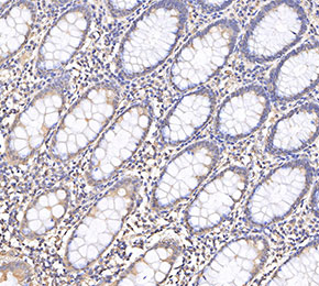 Immunohistochemical analysis of formalin fixed paraffin embedded human colorectal cancer tissue with F3798 at 1:1000 dilution.
使用情報
| Dilution |
|---|
|
| Application |
|---|
| IHC, FCM |
| Source |
|---|
| Mouse Monoclonal Antibody |
| Reactivity |
|---|
| Human, Mouse |
| Storage Buffer |
|---|
| PBS, pH 7.2+50% Glycerol+0.05% BSA+0.01% NaN3 |
| Storage (from the date of receipt) |
|---|
| -20°C (avoid freeze-thaw cycles), 2 years |
| ポジティブコントロール | Human hippocampus; Mouse backskin; SH-SY5Y |
|---|---|
| ネガティブコントロール |
プロトコール
| IHC |
|---|
Experimental Protocol:
Deparaffinization/Rehydration
1. Deparaffinize/hydrate sections:
2. Incubate sections in three washes of xylene for 5 min each.
3. Incubate sections in two washes of 100% ethanol for 10 min each.
4. Incubate sections in two washes of 95% ethanol for 10 min each.
5. Wash sections two times in dH2O for 5 min each.
6.Antigen retrieval: For Citrate: Heat slides in a microwave submersed in 1X citrate unmasking solution until boiling is initiated; continue with 10 min at a sub-boiling temperature (95°-98°C). Cool slides on bench top for 30 min.
Staining
1. Wash sections in dH2O three times for 5 min each.
2. Incubate sections in 3% hydrogen peroxide for 10 min.
3. Wash sections in dH2O two times for 5 min each.
4. Wash sections in wash buffer for 5 min.
5. Block each section with 100–400 µl of blocking solution for 1 hr at room temperature.
6. Remove blocking solution and add 100–400 µl primary antibody diluent in to each section. Incubate overnight at 4°C.
7. Remove antibody solution and wash sections with wash buffer three times for 5 min each.
8. Cover section with 1–3 drops HRPas needed. Incubate in a humidified chamber for 30 min at room temperature.
9. Wash sections three times with wash buffer for 5 min each.
10. Add DAB Chromogen Concentrate to DAB Diluent and mix well before use.
11. Apply 100–400 µl DAB to each section and monitor closely. 1–10 min generally provides an acceptable staining intensity.
12. Immerse slides in dH2O.
13. If desired, counterstain sections with hematoxylin.
14. Wash sections in dH2O two times for 5 min each.
15. Dehydrate sections: Incubate sections in 95% ethanol two times for 10 sec each; Repeat in 100% ethanol, incubating sections two times for 10 sec each; Repeat in xylene, incubating sections two times for 10 sec each.
16. Mount sections with coverslips and mounting medium.
|
生物学的記述
| Specificity |
|---|
CaV1.3 Antibody (Mouse mAb) [G19D14] detects endogenous levels of total CaV1.3 protein. |
| タンパク質の局在 |
|---|
| 細胞内膜系 |
| Uniprot ID |
|---|
| Q01668 |
| Clone |
|---|
| G19D14 |
| Synonym(s) |
|---|
| CACH3, CACN4, CACNL1A2, CCHL1A2, CACNA1D, Voltage-dependent L-type calcium channel subunit alpha-1D, Voltage-gated calcium channel subunit alpha Cav1.3. |
| Background |
|---|
| Cardiac excitation–contraction coupling refers to the sequence of events in which electrical stimulation of a cardiomyocyte triggers muscle contraction in the heart. L-type Ca²⁺ channels are critical to this process, as they facilitate calcium influx and contribute to membrane excitability. Four L-type Ca²⁺ channel subtypes have been identified: Cav1.1, Cav1.2, Cav1.3, and Cav1.4. Cav1.1 is predominantly found in skeletal muscle, while Cav1.4 is mainly expressed in the retina and certain immune cells. Cav1.3 is present in the heart, somatodendritic regions of neurons, endocrine cells, and sensory cells. In the heart, Cav1.3 activity is modulated by multiple neurotransmitters. Phosphorylation by cAMP-dependent protein kinase A (PKA) occurs at serine residues 1743 and 1816 within the C-terminal region. Protein kinase C (PKC) also regulates Cav1.3 in an isozyme-specific manner via phosphorylation at serine 81 in the N-terminal domain. Additionally, alternative splicing within the C-terminus influences channel behavior, notably decreasing Ca²⁺-dependent inactivation. Functionally, Cav1.3 contributes to cardiac pacemaking and atrioventricular (AV) conduction. Dysfunction of Cav1.3 has been associated with sinoatrial node and AV node abnormalities, as well as the development of atrial fibrillation. |
| References |
|---|
技術サポート
ストックの作り方、阻害剤の保管方法、細胞実験や動物実験の際に注意すべき点など、製品を取扱う時に問い合わせが多かった質問に対しては取扱説明書でお答えしています。
他に質問がある場合は、お気軽にお問い合わせください。
* 必須
納期 国内在庫品:受注日の翌日(15時までの受注分) *北海道、九州、沖縄への配送は受注日より2日以上 を要する場合あり 海外在庫品:受注後1〜2週間