
- 阻害剤
- 研究分野別
- PI3K/Akt/mTOR
- Epigenetics
- Methylation
- Immunology & Inflammation
- Protein Tyrosine Kinase
- Angiogenesis
- Apoptosis
- Autophagy
- ER stress & UPR
- JAK/STAT
- MAPK
- Cytoskeletal Signaling
- Cell Cycle
- TGF-beta/Smad
- 化合物ライブラリー
- 抗体
- 新製品
- お問い合わせ

CD59 Antibody (Mouse mAb) [M4G24]
CatNo: F2130
Application:
Reactivity:
-
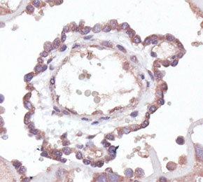 Immunohistochemical analysis of formalin fixed paraffin embedded human placental tissue with F2130 at 1:1000 dilution.
当該製品は品切れ状态で、メールアドレスをご教示いただければ、お客様に返信いたします。
代表番号: 045-509-1970|電子メール:sales@selleck.co.jp
使用情報
| Dilution |
|---|
|
| Application |
|---|
| IHC, FCM |
| Source |
|---|
| Mouse Monoclonal Antibody |
| Reactivity |
|---|
| Human |
| Storage Buffer |
|---|
| PBS, pH 7.2+50% Glycerol+0.05% BSA+0.01% NaN3 |
| Storage (from the date of receipt) |
|---|
| -20°C (avoid freeze-thaw cycles), 2 years |
| Predicted MW |
|---|
| 14 kDa |
| ポジティブコントロール | Human palacenta tissue; Human Peripheral Blood cells |
|---|---|
| ネガティブコントロール |
プロトコール
| IHC |
|---|
Experimental Protocol:
Deparaffinization/Rehydration
1. Deparaffinize/hydrate sections:
2. Incubate sections in three washes of xylene for 5 min each.
3. Incubate sections in two washes of 100% ethanol for 10 min each.
4. Incubate sections in two washes of 95% ethanol for 10 min each.
5. Wash sections two times in dH2O for 5 min each.
6.Antigen retrieval: For Citrate: Heat slides in a microwave submersed in 1X citrate unmasking solution until boiling is initiated; continue with 10 min at a sub-boiling temperature (95°-98°C). Cool slides on bench top for 30 min.
Staining
1. Wash sections in dH2O three times for 5 min each.
2. Incubate sections in 3% hydrogen peroxide for 10 min.
3. Wash sections in dH2O two times for 5 min each.
4. Wash sections in wash buffer for 5 min.
5. Block each section with 100–400 µl of blocking solution for 1 hr at room temperature.
6. Remove blocking solution and add 100–400 µl primary antibody diluent in to each section. Incubate overnight at 4°C.
7. Remove antibody solution and wash sections with wash buffer three times for 5 min each.
8. Cover section with 1–3 drops HRPas needed. Incubate in a humidified chamber for 30 min at room temperature.
9. Wash sections three times with wash buffer for 5 min each.
10. Add DAB Chromogen Concentrate to DAB Diluent and mix well before use.
11. Apply 100–400 µl DAB to each section and monitor closely. 1–10 min generally provides an acceptable staining intensity.
12. Immerse slides in dH2O.
13. If desired, counterstain sections with hematoxylin.
14. Wash sections in dH2O two times for 5 min each.
15. Dehydrate sections: Incubate sections in 95% ethanol two times for 10 sec each; Repeat in 100% ethanol, incubating sections two times for 10 sec each; Repeat in xylene, incubating sections two times for 10 sec each.
16. Mount sections with coverslips and mounting medium.
|
生物学的記述
| Specificity |
|---|
CD59 Antibody (Mouse mAb) [M4G24] detects endogenous levels of total CD59 protein. |
| タンパク質の局在 |
|---|
| 細胞膜、細胞内膜系、細胞外環境 |
| Uniprot ID |
|---|
| P13987 |
| Clone |
|---|
| M4G24 |
| Synonym(s) |
|---|
| CD59, MIC11, MIN1, MIN2, MIN3, MSK21, CD59 glycoprotein, 1F5 antigen, 20 kDa homologous restriction factor, MAC-inhibitory protein, MEM43 antigen, Protectin, HRF-20, HRF20, MAC-IP, MACIF, MIRL |
| Background |
|---|
CD59 is a small, glycosylphosphatidylinositol-anchored membrane glycoprotein that functions as the principal cell-surface inhibitor of the terminal complement pathway, expressed broadly across hematopoietic and non-hematopoietic human cells. The membrane attack complex assembles through the sequential recruitment of complement proteins C5b, C6, C7, C8, and C9, a process in which C8 and C9 each undergo a dramatic conformational transition, unfurling helical bundles within their MACPF domains into membrane-inserting β-hairpins that drive pore formation. CD59 does not bind soluble C8 or C9 but engages both proteins specifically once they are incorporated into the nascent complex, capturing the C5b-8 and C5b-9 assembly intermediates before pore insertion occurs. CD59 binds the pore-forming β-hairpin region of C8, forming an intermolecular β-sheet that physically blocks membrane perforation by C8 itself, while simultaneously deflecting the trajectory of the first incoming C9 β-hairpin as it attempts to insert into the membrane. By rerouting this initial C9 insertion, CD59 restricts the conformational transition required for subsequent C9 monomers to join the growing pore, halting polymerization of the full membrane attack complex rather than blocking any single binding step in isolation. This dual mechanism, direct steric blockade of C8 and indirect disruption of C9 polymerization, allows a single CD59 molecule to arrest membrane attack complex assembly at more than one stage of its stepwise construction. Loss of CD59 from the cell surface, together with the related regulator CD55, results almost universally from somatic mutation in the PIGA gene, which disrupts the first step of glycosylphosphatidylinositol anchor biosynthesis and prevents proper anchoring of both proteins to affected hematopoietic cells. This GPI-anchor deficiency underlies paroxysmal nocturnal hemoglobinuria, in which the resulting absence of CD59 leaves red blood cells unprotected against membrane attack complex assembly, producing chronic complement-mediated intravascular hemolysis. |
| References |
|---|
技術サポート
ストックの作り方、阻害剤の保管方法、細胞実験や動物実験の際に注意すべき点など、製品を取扱う時に問い合わせが多かった質問に対しては取扱説明書でお答えしています。
他に質問がある場合は、お気軽にお問い合わせください。
* 必須

納期 国内在庫品:受注日の翌日(15時までの受注分) *北海道、九州、沖縄への配送は受注日より2日以上 を要する場合あり 海外在庫品:受注後1〜2週間