
- 阻害剤
- 研究分野別
- PI3K/Akt/mTOR
- Epigenetics
- Methylation
- Immunology & Inflammation
- Protein Tyrosine Kinase
- Angiogenesis
- Apoptosis
- Autophagy
- ER stress & UPR
- JAK/STAT
- MAPK
- Cytoskeletal Signaling
- Cell Cycle
- TGF-beta/Smad
- 化合物ライブラリー
- 抗体
- 新製品
- お問い合わせ

CDC42 Antibody (Rabbit mAb) [B14M24]
CatNo: F4204
Application:
Reactivity:
-
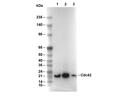 Lane 1: Jurkat, Lane 2: C6, Lane 3: NIH/3T3
当該製品は品切れ状态で、メールアドレスをご教示いただければ、お客様に返信いたします。
代表番号: 045-509-1970|電子メール:sales@selleck.co.jp
キーポイント
WB
転写条件(ウェット): 200 mA, 60 min。
転写条件(ウェット): 200 mA, 60 min。
使用情報
| Dilution |
|---|
|
| Application |
|---|
| WB |
| Source |
|---|
| Rabbit Monoclonal Antibody |
| Reactivity |
|---|
| Human, Mouse, Rat, Bovine |
| Storage Buffer |
|---|
| PBS, pH 7.2+50% Glycerol+0.05% BSA+0.01% NaN3 |
| Storage (from the date of receipt) |
|---|
| -20°C (avoid freeze-thaw cycles), 2 years |
| Predicted MW |
|---|
| 21 kDa |
| ポジティブコントロール | Jurkat cells; C6 cells; NIH/3T3 cells |
|---|---|
| ネガティブコントロール |
プロトコール
| WB |
|---|
Experimental Protocol:
Sample preparation
1. Tissue: Lyse the tissue sample by adding an appropriate volume of ice-cold RIPA/NP-40 Lysis Buffer (containing Protease Inhibitor Cocktail),and homogenize the tissue at a low temperature or lyse it by sonication on ice, then incubate on ice for 30 minutes. 2. Adherent cell: Aspirate the culture medium and wash the cells with ice-cold PBS twice. Lyse the cells by adding an appropriate volume of RIPA/NP-40 Lysis Buffer (containing Protease Inhibitor Cocktail) , sonicate to lyse the cells, and incubate on ice for 30 minutes. 3. Suspension cell: Transfer the culture medium to a pre-cooled centrifuge tube. Centrifuge and aspirate the supernatant. Wash the cells with ice-cold PBS twice. Lyse the cells by adding an appropriate volume of RIPA/NP-40 Lysis Buffer (containing Protease Inhibitor Cocktail) , sonicate to lyse the cells, and incubate on ice for 30 minutes. 4. Place the lysate into a pre-cooled microcentrifuge tube. Centrifuge at 4°C for 15 min. Collect the supernatant;
5. Remove a small volume of lysate to determine the protein concentration;
6. Combine the lysate with protein loading buffer. Boil 20 µL sample under 95-100°C for 5 min. Centrifuge for 5 min after cool down on ice.
Electrophoretic separation
1. According to the concentration of extracted protein, load appropriate amount of protein sample and marker onto SDS-PAGE gels for electrophoresis. Recommended separating gel (lower gel) concentration: 10%. Reference Table for Selecting SDS-PAGE Separation Gel Concentrations 2. Power up 80V for 30 minutes. Then the power supply is adjusted (110 V~150 V), the Marker is observed, and the electrophoresis can be stopped when the indicator band of the predyed protein Marker where the protein is located is properly separated. (Note that the current should not be too large when electrophoresis, too large current (more than 150 mA) will cause the temperature to rise, affecting the result of running glue. If high currents cannot be avoided, an ice bath can be used to cool the bath.)
Transfer membrane
1. Take out the converter, soak the clip and consumables in the pre-cooled converter;
2. Activate PVDF membrane with methanol for 1 min and rinse with transfer buffer;
3. Install it in the order of "black edge of clip - sponge - filter paper - filter paper - glue -PVDF membrane - filter paper - filter paper - sponge - white edge of clip"; 4. The protein was electrotransferred to PVDF membrane. ( 0.45 µm PVDF membrane is recommended ) Reference Table for Selecting PVDF Membrane Pore Size Specifications Recommended conditions for wet transfer: 200 mA, 60 min. ( Note that the transfer conditions can be adjusted according to the protein size. For high-molecular-weight proteins, a higher current and longer transfer time are recommended. However, ensure that the transfer tank remains at a low temperature to prevent gel melting.)
Block
1. After electrotransfer, wash the film with TBST at room temperature for 5 minutes;
2. Incubate the film in the blocking solution for 1 hour at room temperature;
3. Wash the film with TBST for 3 times, 5 minutes each time.
Antibody incubation
1. Use primary antibody dilution buffer to prepare the primary antibody working liquid (recommended dilution ratio for primary antibody 1:1000), gently shake and incubate with the film at 4°C overnight; 2. Wash the film with TBST 3 times, 5 minutes each time;
3. Add the secondary antibody to the blocking solution and incubate with the film gently at room temperature for 1 hour;
4. After incubation, wash the film with TBST 3 times for 5 minutes each time.
Antibody staining
1. Add the prepared ECL luminescent substrate (or select other color developing substrate according to the second antibody) and mix evenly;
2. Incubate with the film for 1 minute, remove excess substrate (keep the film moist), wrap with plastic film, and expose in the imaging system. |
生物学的記述
| Specificity |
|---|
| CDC42 Antibody (Rabbit mAb) [B14M24] detects endogenous levels of total CDC42 protein. |
| タンパク質の局在 |
|---|
| 細胞膜、細胞突起、細胞質 |
| Uniprot ID |
|---|
| P60953 |
| Clone |
|---|
| B14M24 |
| Synonym(s) |
|---|
| CDC42 | CDC42Hs | Cell division control protein 42 homolog | cell division cycle 42 | cell division cycle 42 (GTP binding protein | 25kDa) | dJ224A6.1.1 (cell division cycle 42 (GTP-binding protein | 25kD)) | dJ224A6.1.2 (cell division cycle 42 (GTP-binding protein | 25kD)) | G25K | G25K GTP-binding protein | growth-regulating protein | GTP binding protein | 25kDa | GTP-binding protein | 25kD | small GTP binding protein CDC42 | TKS |
| Background |
|---|
| CDC42 is a small Rho‑family GTPase that acts as a conserved molecular switch controlling actin cytoskeleton organization, cell polarity, membrane trafficking, and signaling pathways that regulate growth and differentiation across diverse mammalian tissues. The protein contains a canonical small GTPase fold with a P‑loop NTP‑binding domain, switch I and II regions that change conformation upon GTP binding and hydrolysis, and a C‑terminal CAAX motif that is prenylated to anchor CDC42 at specific membrane compartments where receptors and scaffolding proteins recruit its dedicated guanine nucleotide exchange factors and GTPase‑activating proteins. CDC42 cycles between GDP‑bound inactive and GTP‑bound active states under the coordinated action of GEFs that catalyze GDP–GTP exchange, GAPs that accelerate intrinsic GTP hydrolysis, and RhoGDIs that bind GDP‑loaded CDC42 and retain it in the cytosol until appropriate signals release it for membrane targeting and activation. Active CDC42 engages multiple effector proteins, including p21‑activated kinases, N‑WASP/WASP, and PAR polarity complexes, and these effectors connect CDC42 activation to Arp2/3‑dependent actin nucleation, formation of actin‑rich filopodia, establishment of apical–basal polarity, and organization of adherens and tight junctions, which coordinates epithelial morphogenesis, directed migration, and lumen and barrier formation. CDC42 also interacts with IQGAP proteins, MRCK kinases, and regulators at the Golgi and plasma membrane to integrate actomyosin contractility with vesicle trafficking and microtubule dynamics, shaping junctional stability, polarized secretion, and organelle positioning. In endocrine tissues, glucose‑stimulated CDC42 activation functions as an early signal in stimulus–secretion coupling for insulin, where CDC42 regulates cortical actin remodeling and recruitment of exocytotic machinery required for the sustained second phase of insulin release. In signaling networks controlling proliferation and survival, CDC42 modulates pathways such as EGFR–mTORC1 and contributes to activation of downstream cascades including MAPK and PI3K, which influence transcriptional programs and cell‑cycle progression. Germline mutations and dysregulated CDC42 activity associate with developmental and autoinflammatory syndromes, while overexpression or hyperactivation in tumors correlates with enhanced motility, invasive behavior, and metastatic dissemination. |
| References |
|---|
技術サポート
ストックの作り方、阻害剤の保管方法、細胞実験や動物実験の際に注意すべき点など、製品を取扱う時に問い合わせが多かった質問に対しては取扱説明書でお答えしています。
他に質問がある場合は、お気軽にお問い合わせください。
* 必須

納期 国内在庫品:受注日の翌日(15時までの受注分) *北海道、九州、沖縄への配送は受注日より2日以上 を要する場合あり 海外在庫品:受注後1〜2週間