
- 阻害剤
- 研究分野別
- PI3K/Akt/mTOR
- Epigenetics
- Methylation
- Immunology & Inflammation
- Protein Tyrosine Kinase
- Angiogenesis
- Apoptosis
- Autophagy
- ER stress & UPR
- JAK/STAT
- MAPK
- Cytoskeletal Signaling
- Cell Cycle
- TGF-beta/Smad
- 化合物ライブラリー
- 抗体
- 新製品
- お問い合わせ
Collagen VII Antibody (Mouse mAb) [K3G14]
Catalog No.: F3214
Application:
Reactivity:
-
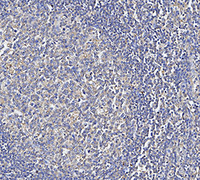 Immunohistochemical analysis of formalin fixed paraffin embedded human tonsils tissue with F3214 at 1:500 dilution.
使用情報
| Dilution |
|---|
|
| Application |
|---|
| IHC-Fr |
| Source |
|---|
| Mouse Monoclonal Antibody |
| Reactivity |
|---|
| Human |
| Storage Buffer |
|---|
| PBS, pH 7.2+50% Glycerol+0.05% BSA+0.01% NaN3 |
| Storage (from the date of receipt) |
|---|
| -20°C (avoid freeze-thaw cycles), 2 years |
| ポジティブコントロール | Human tonsil |
|---|---|
| ネガティブコントロール |
プロトコール
| IHC |
|---|
Experimental Protocol:
Deparaffinization/Rehydration
1. Deparaffinize/hydrate sections:
2. Incubate sections in three washes of xylene for 5 min each.
3. Incubate sections in two washes of 100% ethanol for 10 min each.
4. Incubate sections in two washes of 95% ethanol for 10 min each.
5. Wash sections two times in dH2O for 5 min each.
6.Antigen retrieval: For Citrate: Heat slides in a microwave submersed in 1X citrate unmasking solution until boiling is initiated; continue with 10 min at a sub-boiling temperature (95°-98°C). Cool slides on bench top for 30 min.
Staining
1. Wash sections in dH2O three times for 5 min each.
2. Incubate sections in 3% hydrogen peroxide for 10 min.
3. Wash sections in dH2O two times for 5 min each.
4. Wash sections in wash buffer for 5 min.
5. Block each section with 100–400 µl of blocking solution for 1 hr at room temperature.
6. Remove blocking solution and add 100–400 µl primary antibody diluent in to each section. Incubate overnight at 4°C.
7. Remove antibody solution and wash sections with wash buffer three times for 5 min each.
8. Cover section with 1–3 drops HRPas needed. Incubate in a humidified chamber for 30 min at room temperature.
9. Wash sections three times with wash buffer for 5 min each.
10. Add DAB Chromogen Concentrate to DAB Diluent and mix well before use.
11. Apply 100–400 µl DAB to each section and monitor closely. 1–10 min generally provides an acceptable staining intensity.
12. Immerse slides in dH2O.
13. If desired, counterstain sections with hematoxylin.
14. Wash sections in dH2O two times for 5 min each.
15. Dehydrate sections: Incubate sections in 95% ethanol two times for 10 sec each; Repeat in 100% ethanol, incubating sections two times for 10 sec each; Repeat in xylene, incubating sections two times for 10 sec each.
16. Mount sections with coverslips and mounting medium.
|
生物学的記述
| Specificity |
|---|
Collagen VII Antibody (Mouse mAb) [K3G14] detects endogenous levels of total Collagen VII protein. |
| タンパク質の局在 |
|---|
| 基底膜、細胞外マトリックス、細胞外環境 |
| Uniprot ID |
|---|
| Q02388 |
| Clone |
|---|
| K3G14 |
| Synonym(s) |
|---|
| Collagen alpha-1(VII) chain, Long-chain collagen, LC collagen, COL7A1 |
| Background |
|---|
| Collagen VII is a specialized fibrillar collagen primarily responsible for forming anchoring fibrils that stabilize the dermal-epidermal junction in the skin. It is encoded by the COL7A1 gene and is expressed predominantly in epidermal keratinocytes and dermal fibroblasts. Structurally, collagen VII is a homotrimer composed of three pro-α1(VII) chains, each featuring a central triple-helical domain (~1,530 amino acids) interrupted by 19 imperfections, including a protease-sensitive 39-amino acid “hinge” region. This central domain is flanked by two non-collagenous domains: NC-1 at the N-terminus, which contains adhesive modules like fibronectin type III repeats and a von Willebrand factor A domain, and NC-2 at the C-terminus, which includes a Kunitz protease inhibitor-like segment. Two collagen VII molecules assemble into antiparallel dimers stabilized by disulfide bonds and further aggregate into anchoring fibrils. Functionally, collagen VII anchors the basement membrane to the underlying dermis by binding with high affinity to basement membrane components such as laminin-332 and collagen IV, thereby maintaining skin integrity; its mutations cause dystrophic epidermolysis bullosa (DEB), a severe blistering skin disorders. |
| References |
|---|
技術サポート
ストックの作り方、阻害剤の保管方法、細胞実験や動物実験の際に注意すべき点など、製品を取扱う時に問い合わせが多かった質問に対しては取扱説明書でお答えしています。
他に質問がある場合は、お気軽にお問い合わせください。
* 必須
納期 国内在庫品:受注日の翌日(15時までの受注分) *北海道、九州、沖縄への配送は受注日より2日以上 を要する場合あり 海外在庫品:受注後1〜2週間