
- 阻害剤
- 研究分野別
- PI3K/Akt/mTOR
- Epigenetics
- Methylation
- Immunology & Inflammation
- Protein Tyrosine Kinase
- Angiogenesis
- Apoptosis
- Autophagy
- ER stress & UPR
- JAK/STAT
- MAPK
- Cytoskeletal Signaling
- Cell Cycle
- TGF-beta/Smad
- 化合物ライブラリー
- 抗体
- 新製品
- お問い合わせ

DLL4 Antibody (Hamster mAb) [N12K10]
CatNo: F5840
Application:
Reactivity:
-
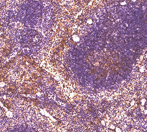 Immunohistochemical analysis of formalin fixed paraffin embedded mouse spleen tissue with F5840 at 1:100 dilution.
当該製品は品切れ状态で、メールアドレスをご教示いただければ、お客様に返信いたします。
代表番号: 045-509-1970|電子メール:sales@selleck.co.jp
使用情報
| Dilution |
|---|
|
| Application |
|---|
| IHC, FCM |
| Source |
|---|
| Hamster Monoclonal Antibody |
| Reactivity |
|---|
| Mouse |
| Storage Buffer |
|---|
| PBS, pH 7.2+50% Glycerol+0.05% BSA+0.01% NaN3 |
| Storage (from the date of receipt) |
|---|
| -20°C (avoid freeze-thaw cycles), 2 years |
| Predicted MW |
|---|
| 75 kDa |
プロトコール
| IHC |
|---|
Experimental Protocol:
Deparaffinization/Rehydration
1. Deparaffinize/hydrate sections:
2. Incubate sections in three washes of xylene for 5 min each.
3. Incubate sections in two washes of 100% ethanol for 10 min each.
4. Incubate sections in two washes of 95% ethanol for 10 min each.
5. Wash sections two times in dH2O for 5 min each.
6.Antigen retrieval: For Citrate: Heat slides in a microwave submersed in 1X citrate unmasking solution until boiling is initiated; continue with 10 min at a sub-boiling temperature (95°-98°C). Cool slides on bench top for 30 min.
Staining
1. Wash sections in dH2O three times for 5 min each.
2. Incubate sections in 3% hydrogen peroxide for 10 min.
3. Wash sections in dH2O two times for 5 min each.
4. Wash sections in wash buffer for 5 min.
5. Block each section with 100–400 µl of blocking solution for 1 hr at room temperature.
6. Remove blocking solution and add 100–400 µl primary antibody diluent in to each section. Incubate overnight at 4°C.
7. Remove antibody solution and wash sections with wash buffer three times for 5 min each.
8. Cover section with 1–3 drops HRPas needed. Incubate in a humidified chamber for 30 min at room temperature.
9. Wash sections three times with wash buffer for 5 min each.
10. Add DAB Chromogen Concentrate to DAB Diluent and mix well before use.
11. Apply 100–400 µl DAB to each section and monitor closely. 1–10 min generally provides an acceptable staining intensity.
12. Immerse slides in dH2O.
13. If desired, counterstain sections with hematoxylin.
14. Wash sections in dH2O two times for 5 min each.
15. Dehydrate sections: Incubate sections in 95% ethanol two times for 10 sec each; Repeat in 100% ethanol, incubating sections two times for 10 sec each; Repeat in xylene, incubating sections two times for 10 sec each.
16. Mount sections with coverslips and mounting medium.
|
生物学的記述
| Specificity |
|---|
DLL4 Antibody (Hamster mAb) [N12K10] detects endogenous levels of total δ-Like Protein 4 protein. |
| タンパク質の局在 |
|---|
| 細胞膜、細胞内膜系 |
| Uniprot ID |
|---|
| Q9JI71 |
| Clone |
|---|
| N12K10 |
| Synonym(s) |
|---|
| DLL4,AOS6 |
| Background |
|---|
Delta‑like protein 4 (DLL4) is an endothelial‑enriched Notch ligand of the Delta/Serrate/Lag family that presents an extracellular DSL motif and tandem EGF‑like repeats to engage Notch receptors on adjacent cells, where ligand‑induced receptor cleavage generates the Notch intracellular domain and drives transcriptional programs that sculpt vascular patterning. DLL4 expression is largely restricted to arterial and actively sprouting endothelium, with highest levels in tip and stalk cells of angiogenic vessels, and is strongly induced by pro‑angiogenic cues such as VEGF and hypoxia‑regulated HIF‑1α, placing DLL4/Notch signaling downstream of VEGF receptor activation in the hierarchy of angiogenic control. At the sprouting front, DLL4 expressed on emerging tip cells activates Notch in neighboring stalk cells, down‑regulates VEGF receptor expression, limits excessive tip‑cell formation, and enforces a hierarchical branching pattern that yields a functional, perfused vascular network; genetic or pharmacologic blockade of DLL4/Notch signaling disrupts this lateral inhibition, increases endothelial proliferation and branching, and produces dense but poorly perfused, non‑productive vasculature. DLL4 is up‑regulated in the vasculature of many solid cancers, including colorectal, breast, renal, pancreatic, and glioblastoma models, and that forced DLL4 expression in tumor or stromal compartments reduces vessel density but enlarges lumen size and improves vascular function, lowering hypoxia and apoptosis and supporting tumor growth, whereas soluble DLL4 decoys or anti‑DLL4 antibodies increase vessel number yet impair perfusion and suppress tumor expansion, including in settings that are resistant to anti‑VEGF therapy. Across preclinical models, high DLL4 levels in tumor endothelium correlate with more mature vasculature and shorter patient survival, while DLL4 blockade consistently drives hyper‑sprouting and functional collapse of the tumor vasculature, identifying DLL4/Notch as a central negative regulator of angiogenic output that modulates and intersects with VEGF signaling rather than simply mirroring it. |
| References |
|---|
技術サポート
ストックの作り方、阻害剤の保管方法、細胞実験や動物実験の際に注意すべき点など、製品を取扱う時に問い合わせが多かった質問に対しては取扱説明書でお答えしています。
他に質問がある場合は、お気軽にお問い合わせください。
* 必須

納期 国内在庫品:受注日の翌日(15時までの受注分) *北海道、九州、沖縄への配送は受注日より2日以上 を要する場合あり 海外在庫品:受注後1〜2週間