
- 阻害剤
- 研究分野別
- PI3K/Akt/mTOR
- Epigenetics
- Methylation
- Immunology & Inflammation
- Protein Tyrosine Kinase
- Angiogenesis
- Apoptosis
- Autophagy
- ER stress & UPR
- JAK/STAT
- MAPK
- Cytoskeletal Signaling
- Cell Cycle
- TGF-beta/Smad
- 化合物ライブラリー
- 抗体
- 新製品
- お問い合わせ

DMT1/SLC11A2 Antibody (Rabbit mAb) [C16J1]
CatNo: F5070
Application:
Reactivity:
-
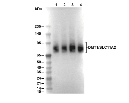 Lane 1: Caco-2, Lane 2: Huh7, Lane 3: 293T, Lane 4: SH-SY5Y
使用情報
| Dilution |
|---|
|
| Application |
|---|
| WB, IP |
| Source |
|---|
| Rabbit Monoclonal Antibody |
| Reactivity |
|---|
| Human |
| Storage Buffer |
|---|
| PBS, pH 7.2+50% Glycerol+0.05% BSA+0.01% NaN3 |
| Storage (from the date of receipt) |
|---|
| -20°C (avoid freeze-thaw cycles), 2 years |
| Predicted MW |
|---|
| 62 kDa |
| ポジティブコントロール | SK-MEL-5 cells; SK-MEL-2 cells |
|---|---|
| ネガティブコントロール |
プロトコール
| WB |
|---|
Experimental Protocol:
Sample preparation
1. Tissue: Lyse the tissue sample by adding an appropriate volume of ice-cold RIPA/NP-40 Lysis Buffer (containing Protease Inhibitor Cocktail),and homogenize the tissue at a low temperature or lyse it by sonication on ice, then incubate on ice for 30 minutes. 2. Adherent cell: Aspirate the culture medium and wash the cells with ice-cold PBS twice. Lyse the cells by adding an appropriate volume of RIPA/NP-40 Lysis Buffer (containing Protease Inhibitor Cocktail) , sonicate to lyse the cells, and incubate on ice for 30 minutes. 3. Suspension cell: Transfer the culture medium to a pre-cooled centrifuge tube. Centrifuge and aspirate the supernatant. Wash the cells with ice-cold PBS twice. Lyse the cells by adding an appropriate volume of RIPA/NP-40 Lysis Buffer (containing Protease Inhibitor Cocktail) , sonicate to lyse the cells, and incubate on ice for 30 minutes. 4. Place the lysate into a pre-cooled microcentrifuge tube. Centrifuge at 4°C for 15 min. Collect the supernatant;
5. Remove a small volume of lysate to determine the protein concentration;
6. Combine the lysate with protein loading buffer. Boil 20 µL sample under 95-100°C for 5 min. Centrifuge for 5 min after cool down on ice.
Electrophoretic separation
1. According to the concentration of extracted protein, load appropriate amount of protein sample and marker onto SDS-PAGE gels for electrophoresis. Recommended separating gel (lower gel) concentration: 10%. Reference Table for Selecting SDS-PAGE Separation Gel Concentrations 2. Power up 80V for 30 minutes. Then the power supply is adjusted (110 V~150 V), the Marker is observed, and the electrophoresis can be stopped when the indicator band of the predyed protein Marker where the protein is located is properly separated. (Note that the current should not be too large when electrophoresis, too large current (more than 150 mA) will cause the temperature to rise, affecting the result of running glue. If high currents cannot be avoided, an ice bath can be used to cool the bath.)
Transfer membrane
1. Take out the converter, soak the clip and consumables in the pre-cooled converter;
2. Activate PVDF membrane with methanol for 1 min and rinse with transfer buffer;
3. Install it in the order of "black edge of clip - sponge - filter paper - filter paper - glue -PVDF membrane - filter paper - filter paper - sponge - white edge of clip"; 4. The protein was electrotransferred to PVDF membrane. ( 0.45 µm PVDF membrane is recommended ) Reference Table for Selecting PVDF Membrane Pore Size Specifications Recommended conditions for wet transfer: 200 mA, 120 min. ( Note that the transfer conditions can be adjusted according to the protein size. For high-molecular-weight proteins, a higher current and longer transfer time are recommended. However, ensure that the transfer tank remains at a low temperature to prevent gel melting.)
Block
1. After electrotransfer, wash the film with TBST at room temperature for 5 minutes;
2. Incubate the film in the blocking solution for 1 hour at room temperature;
3. Wash the film with TBST for 3 times, 5 minutes each time.
Antibody incubation
1. Use primary antibody dilution buffer to prepare the primary antibody working liquid (recommended dilution ratio for primary antibody 1:1000), gently shake and incubate with the film at 4°C overnight; 2. Wash the film with TBST 3 times, 5 minutes each time;
3. Add the secondary antibody to the blocking solution and incubate with the film gently at room temperature for 1 hour;
4. After incubation, wash the film with TBST 3 times for 5 minutes each time.
Antibody staining
1. Add the prepared ECL luminescent substrate (or select other color developing substrate according to the second antibody) and mix evenly;
2. Incubate with the film for 1 minute, remove excess substrate (keep the film moist), wrap with plastic film, and expose in the imaging system. |
生物学的記述
| Specificity |
|---|
| DMT1/SLC11A2 Antibody (Rabbit mAb) [C16J1] detects endogenous levels of total DMT1/SLC11A2 protein. |
| タンパク質の局在 |
|---|
| 細胞膜、エンドソーム、ゴルジ装置、リソソーム、細胞内膜系、ミトコンドリア |
| Uniprot ID |
|---|
| P49281 |
| Clone |
|---|
| C16J1 |
| Synonym(s) |
|---|
| Natural resistance-associated macrophage protein 2, NRAMP 2, Divalent cation transporter 1, Divalent metal transporter 1 (DMT-1), Solute carrier family 11 member 2, SLC11A2, DCT1, DMT1, NRAMP2 |
| Background |
|---|
| DMT1 (SLC11A2/NRAMP2) is a proton‑coupled divalent metal transporter of the SLC11 family that provides the major route for non‑heme iron entry into enterocytes and for transferrin‑derived iron exit from endosomes in erythroid precursors and other cells, thereby linking luminal, endosomal, and cytosolic iron pools to systemic iron homeostasis and intracellular metal balance. The polytopic transporter spans the membrane with multiple transmembrane helices that form a substrate translocation pathway and couple inward movement of Fe²⁺ and other divalent cations to the proton gradient, operating on the apical brush‑border membrane of duodenal enterocytes to import Fe²⁺ released from dietary sources and on endosomal membranes in erythroblasts to move Fe²⁺ from acidified transferrin‑containing endosomes into the cytosol for heme synthesis. Four main DMT1 isoforms arise from alternative promoter usage and 3′ UTR splicing, differing in N‑terminal sequence and presence or absence of an iron‑responsive element (IRE) in the 3′ UTR; these isoforms show distinct tissue and subcellular distributions and are differentially regulated by systemic iron status via HIF2α‑dependent transcription and IRE/IRP‑mediated post‑transcriptional control, allowing DMT1 expression to increase in iron deficiency and decrease when iron stores are replete. Functionally, DMT1 is selective for divalent cations and transports Fe²⁺ with high preference but also mediates uptake of Mn²⁺, Co²⁺, Cd²⁺, and other metals, which makes it central not only to physiological iron absorption and erythropoiesis but also to manganese handling and potential accumulation of toxic metals under environmental or therapeutic exposure. Biallelic loss‑of‑function mutations in SLC11A2 cause an ultra‑rare hypochromic microcytic anemia with iron overload (AHMIO1), where impaired DMT1 activity in erythroblasts reduces endosomal iron delivery to the cytosol, producing iron‑restricted erythropoiesis despite high serum iron, while compensatory upregulation of intestinal DMT1 and continued iron absorption promote hepatic iron overload, leading to anemia with paradoxically elevated serum iron and liver dysfunction. Missense mutations such as G75R and N491S impair DMT1 trafficking and stability, causing mislocalization to lysosomes, reduced transporter levels in patient lymphoblasts, and diminished Fe²⁺ transport, and clinical reports indicate that recombinant erythropoietin can improve the anemic phenotype and may help mobilize hepatic iron in such patients, making SLC11A2 a mechanistically informative genetic cause of combined anemia and iron overload. In the nervous system, DMT1 expression is detected in dopaminergic neurons of the substantia nigra, and the IRE‑containing isoform (DMT1+IRE) is upregulated in toxin‑based Parkinson’s disease models and in postmortem PD nigra, where increased DMT1 correlates with elevated nigral iron, enhanced Fe²⁺ uptake, reactive oxygen species generation, and oxidative stress; iron chelation attenuates these effects, supporting a pathogenic role for DMT1‑mediated iron accumulation in nigral neurodegeneration. |
| References |
|---|
技術サポート
ストックの作り方、阻害剤の保管方法、細胞実験や動物実験の際に注意すべき点など、製品を取扱う時に問い合わせが多かった質問に対しては取扱説明書でお答えしています。
他に質問がある場合は、お気軽にお問い合わせください。
* 必須

納期 国内在庫品:受注日の翌日(15時までの受注分) *北海道、九州、沖縄への配送は受注日より2日以上 を要する場合あり 海外在庫品:受注後1〜2週間