
- 阻害剤
- 研究分野別
- PI3K/Akt/mTOR
- Epigenetics
- Methylation
- Immunology & Inflammation
- Protein Tyrosine Kinase
- Angiogenesis
- Apoptosis
- Autophagy
- ER stress & UPR
- JAK/STAT
- MAPK
- Cytoskeletal Signaling
- Cell Cycle
- TGF-beta/Smad
- 化合物ライブラリー
- 抗体
- 新製品
- お問い合わせ

GABARAPL2/GATE-16 Antibody (Rabbit mAb) [J14F14]
Catalog No.: F5034
Application:
Reactivity:
-
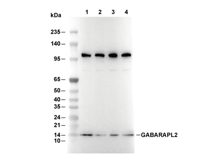 Lane 1: A20, Lane 2: COS-7, Lane 3: RD, Lane 4: C6
当該製品は品切れ状态で、メールアドレスをご教示いただければ、お客様に返信いたします。
代表番号: 045-509-1970|電子メール:sales@selleck.co.jp
キーポイント
WB
SDS-PAGE の分離ゲルの推奨濃度:20%。
転写条件(ウェット): 200 mA, 60 min,Recommended to use 0.22 μm PVDF 膜の使用をお勧めします。
SDS-PAGE の分離ゲルの推奨濃度:20%。
転写条件(ウェット): 200 mA, 60 min,Recommended to use 0.22 μm PVDF 膜の使用をお勧めします。
使用情報
| Dilution |
|---|
|
| Application |
|---|
| WB |
| Source |
|---|
| Rabbit Monoclonal Antibody |
| Reactivity |
|---|
| Human, Mouse, Rat, Monkey |
| Storage Buffer |
|---|
| PBS, pH 7.2+50% Glycerol+0.05% BSA+0.01% NaN3 |
| Storage (from the date of receipt) |
|---|
| -20°C (avoid freeze-thaw cycles), 2 years |
| Predicted MW |
|---|
| 14 kDa |
| ポジティブコントロール | A172 cells (chloroquine, 50 μM, overnight); HeLa cells (chloroquine, 50 μM, overnight); RL cells; RPMI 8226 cells; LN18 cells; RD cells; A20 cells; C6 cells; H-4-II-E cells; COS-7 cells |
|---|---|
| ネガティブコントロール | HeLa cells; A172 cells |
プロトコール
| WB |
|---|
Experimental Protocol:
Sample preparation
1. Tissue: Lyse the tissue sample by adding an appropriate volume of ice-cold RIPA/NP-40 Lysis Buffer (containing Protease Inhibitor Cocktail),and homogenize the tissue at a low temperature or lyse it by sonication on ice, then incubate on ice for 30 minutes. 2. Adherent cell: Aspirate the culture medium and wash the cells with ice-cold PBS twice. Lyse the cells by adding an appropriate volume of RIPA/NP-40 Lysis Buffer (containing Protease Inhibitor Cocktail) , sonicate to lyse the cells, and incubate on ice for 30 minutes. 3. Suspension cell: Transfer the culture medium to a pre-cooled centrifuge tube. Centrifuge and aspirate the supernatant. Wash the cells with ice-cold PBS twice. Lyse the cells by adding an appropriate volume of RIPA/NP-40 Lysis Buffer (containing Protease Inhibitor Cocktail) , sonicate to lyse the cells, and incubate on ice for 30 minutes. 4. Place the lysate into a pre-cooled microcentrifuge tube. Centrifuge at 4°C for 15 min. Collect the supernatant;
5. Remove a small volume of lysate to determine the protein concentration;
6. Combine the lysate with protein loading buffer. Boil 20 µL sample under 95-100°C for 5 min. Centrifuge for 5 min after cool down on ice.
Electrophoretic separation
1. According to the concentration of extracted protein, load appropriate amount of protein sample and marker onto SDS-PAGE gels for electrophoresis. Recommended separating gel (lower gel) concentration: 20%. Reference Table for Selecting SDS-PAGE Separation Gel Concentrations 2. Power up 80V for 30 minutes. Then the power supply is adjusted (110 V~150 V), the Marker is observed, and the electrophoresis can be stopped when the indicator band of the predyed protein Marker where the protein is located is properly separated. (Note that the current should not be too large when electrophoresis, too large current (more than 150 mA) will cause the temperature to rise, affecting the result of running glue. If high currents cannot be avoided, an ice bath can be used to cool the bath.)
Transfer membrane
1. Take out the converter, soak the clip and consumables in the pre-cooled converter;
2. Activate PVDF membrane with methanol for 1 min and rinse with transfer buffer;
3. Install it in the order of "black edge of clip - sponge - filter paper - filter paper - glue -PVDF membrane - filter paper - filter paper - sponge - white edge of clip"; 4. The protein was electrotransferred to PVDF membrane. ( 0.22 µm PVDF membrane is recommended )) Reference Table for Selecting PVDF Membrane Pore Size Specifications Recommended conditions for wet transfer: 200 mA, 60 min. ( Note that the transfer conditions can be adjusted according to the protein size. For high-molecular-weight proteins, a higher current and longer transfer time are recommended. However, ensure that the transfer tank remains at a low temperature to prevent gel melting.)
Block
1. After electrotransfer, wash the film with TBST at room temperature for 5 minutes;
2. Incubate the film in the blocking solution for 1 hour at room temperature;
3. Wash the film with TBST for 3 times, 5 minutes each time.
Antibody incubation
1. Use 5% skim milk powder to prepare the primary antibody working liquid (recommended dilution ratio for primary antibody 1:1000), gently shake and incubate with the film at 4°C overnight; 2. Wash the film with TBST 3 times, 5 minutes each time;
3. Add the secondary antibody to the blocking solution and incubate with the film gently at room temperature for 1 hour;
4. After incubation, wash the film with TBST 3 times for 5 minutes each time.
Antibody staining
1. Add the prepared ECL luminescent substrate (or select other color developing substrate according to the second antibody) and mix evenly;
2. Incubate with the film for 1 minute, remove excess substrate (keep the film moist), wrap with plastic film, and expose in the imaging system. |
生物学的記述
| Specificity |
|---|
| GABARAPL2/GATE-16 Antibody (Rabbit mAb) [J14F14] detects endogenous levels of total GABARAPL2/GATE-16 protein. |
| タンパク質の局在 |
|---|
| 細胞質小胞、小胞体、ゴルジ装置、細胞内膜系 |
| Uniprot ID |
|---|
| P60520 |
| Clone |
|---|
| J14F14 |
| Synonym(s) |
|---|
| ATG8; ATG8C; FLC3A; GABA type A receptor associated protein like 2; GATE-16; GATE16; GBRL2; GEF-2; GEF2; MAP1 light chain 3 related protein; MAP1 light chain 3-related protein |
| Background |
|---|
| GABARAPL2, also known as GATE-16, belongs to the mammalian Atg8 protein family and specifically functions within the GABARAP subfamily, which is distinguished from the LC3 subfamily by unique interaction partners and stage-specific roles in autophagy. The protein adopts a ubiquitin-like fold and undergoes post-translational processing wherein the cysteine protease ATG4 cleaves the carboxyl terminus to expose a glycine residue, which then becomes conjugated to phosphatidylethanolamine on autophagosomal membranes through a cascade involving the E1-like enzyme ATG7 and the E2-like enzyme ATG3. GABARAPL2 orchestrates multiple distinct cellular pathways through its capacity to interact with diverse protein partners, functioning both within and beyond conventional autophagy. The protein directly binds N-ethylmaleimide-sensitive factor (NSF) and stimulates its ATPase activity, which subsequently enhances NSF association with the Golgi v-SNARE GOS-28, thereby coupling NSF enzymatic function with SNARE activation to regulate intra-Golgi membrane transport. Within the autophagy pathway, GABARAPL2 operates at a temporally distinct phase compared to LC3 proteins—while LC3 family members facilitate early phagophore membrane elongation, GABARAPL2 and its subfamily members execute critical functions during late-stage autophagosome maturation and fusion with lysosomes, ensuring cargo clearance. GABARAPL2 associates with cargo receptors through LC3-interacting region (LIR) motifs on partner proteins, including the selective autophagy receptor p62/SQSTM1, enabling targeted degradation of ubiquitinated protein aggregates. The protein exhibits specialized functionality in mitophagy, maintaining mitochondrial quality control through selective elimination of damaged mitochondria, which regulates cellular energy homeostasis and prevents excessive reactive oxygen species accumulation. Beyond autophagy, GABARAPL2 mediates antimicrobial defense by specifically associating with the small GTPase ADP-ribosylation factor 1 (Arf1) to promote uniform cytosolic distribution of interferon-inducible GTPases, and loss of GABARAPL2 impairs Arf1 activation, causing interferon-inducible GTPase aggregation and compromising clearance of intracellular pathogens such as Toxoplasma. The protein further participates in endoplasmic reticulum remodeling through interaction with the reticulophagy receptor TEX264, facilitating ER subdomain conversion into autophagosomes during nutrient stress. GABARAPL2 also localizes at the ER through binding the long-chain-fatty-acid-CoA ligase ACSL3, anchoring the ubiquitin-like modifier activating enzyme UBA5 and functionally coupling lipid droplet biogenesis with UFM1-mediated ufmylation pathways. Additionally, GABARAPL2 activates the CUL3(KBTBD6/7) E3 ubiquitin ligase complex locally, promoting ubiquitination and degradation of TIAM1, a guanyl-nucleotide exchange factor for RAC1, thereby influencing cytoskeletal organization, cell migration, and proliferation. Dysregulation of GABARAPL2 expression or function associates with impaired autophagy flux and defective antimicrobial responses, and the protein's multifunctional nature positions it as a regulatory hub integrating membrane trafficking, autophagy, immune defense, and metabolic signaling. |
| References |
|---|
技術サポート
ストックの作り方、阻害剤の保管方法、細胞実験や動物実験の際に注意すべき点など、製品を取扱う時に問い合わせが多かった質問に対しては取扱説明書でお答えしています。
他に質問がある場合は、お気軽にお問い合わせください。
* 必須

納期 国内在庫品:受注日の翌日(15時までの受注分) *北海道、九州、沖縄への配送は受注日より2日以上 を要する場合あり 海外在庫品:受注後1〜2週間