
- 阻害剤
- 研究分野別
- PI3K/Akt/mTOR
- Epigenetics
- Methylation
- Immunology & Inflammation
- Protein Tyrosine Kinase
- Angiogenesis
- Apoptosis
- Autophagy
- ER stress & UPR
- JAK/STAT
- MAPK
- Cytoskeletal Signaling
- Cell Cycle
- TGF-beta/Smad
- 化合物ライブラリー
- 抗体
- 新製品
- お問い合わせ

IFN-γ Antibody (Rabbit mAb) [A12D6]
CatNo: F6371
Application:
Reactivity:
-
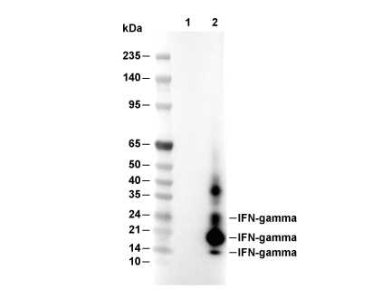 Lane 1: 293T (mock transfected), Lane 2: 293T (mIFN-γ transfected)
当該製品は品切れ状态で、メールアドレスをご教示いただければ、お客様に返信いたします。
代表番号: 045-509-1970|電子メール:sales@selleck.co.jp
キーポイント
WB
転写条件(ウェット): 200 mA, 60 min, 0.22 μm PVDF 膜の使用をお勧めします。
転写条件(ウェット): 200 mA, 60 min, 0.22 μm PVDF 膜の使用をお勧めします。
使用情報
| Dilution |
|---|
|
| Application |
|---|
| WB |
| Source |
|---|
| Rabbit Monoclonal Antibody |
| Reactivity |
|---|
| Mouse |
| Storage Buffer |
|---|
| PBS, pH 7.2+50% Glycerol+0.05% BSA+0.01% NaN3 |
| Storage (from the date of receipt) |
|---|
| -20°C (avoid freeze-thaw cycles), 2 years |
| Predicted MW Observed MW |
|---|
| 18 kDa 17 kDa, 19 kDa, 23 kDa |
| *なぜ予測分子量と実際の分子量が異なるのか? 下記の原因により、実際の分子量が予測と異なる:タンパク質の翻訳後修飾(リン酸化/糖鎖付加),スプライシングバリアント,イソフォーム,相対的な電荷,ポリマー。 |
| ポジティブコントロール | CTLL-2 cells (TPA, 80 nM, 5 h; Ionomycin, Calcium Salt, 3 μM, 5 h; Brefeldin A, 300 ng/mL, last 4 h of stimulation) |
|---|---|
| ネガティブコントロール | CTLL-2 cells |
プロトコール
| WB |
|---|
Experimental Protocol:
Sample preparation
1. Tissue: Lyse the tissue sample by adding an appropriate volume of ice-cold RIPA/NP-40 Lysis Buffer (containing Protease Inhibitor Cocktail),and homogenize the tissue at a low temperature or lyse it by sonication on ice, then incubate on ice for 30 minutes. 2. Adherent cell: Aspirate the culture medium and wash the cells with ice-cold PBS twice. Lyse the cells by adding an appropriate volume of RIPA/NP-40 Lysis Buffer (containing Protease Inhibitor Cocktail) , sonicate to lyse the cells, and incubate on ice for 30 minutes. 3. Suspension cell: Transfer the culture medium to a pre-cooled centrifuge tube. Centrifuge and aspirate the supernatant. Wash the cells with ice-cold PBS twice. Lyse the cells by adding an appropriate volume of RIPA/NP-40 Lysis Buffer (containing Protease Inhibitor Cocktail) , sonicate to lyse the cells, and incubate on ice for 30 minutes. 4. Place the lysate into a pre-cooled microcentrifuge tube. Centrifuge at 4°C for 15 min. Collect the supernatant;
5. Remove a small volume of lysate to determine the protein concentration;
6. Combine the lysate with protein loading buffer. Boil 20 µL sample under 95-100°C for 5 min. Centrifuge for 5 min after cool down on ice.
Electrophoretic separation
1. According to the concentration of extracted protein, load appropriate amount of protein sample and marker onto SDS-PAGE gels for electrophoresis. Recommended separating gel (lower gel) concentration: 10%. Reference Table for Selecting SDS-PAGE Separation Gel Concentrations 2. Power up 80V for 30 minutes. Then the power supply is adjusted (110 V~150 V), the Marker is observed, and the electrophoresis can be stopped when the indicator band of the predyed protein Marker where the protein is located is properly separated. (Note that the current should not be too large when electrophoresis, too large current (more than 150 mA) will cause the temperature to rise, affecting the result of running glue. If high currents cannot be avoided, an ice bath can be used to cool the bath.)
Transfer membrane
1. Take out the converter, soak the clip and consumables in the pre-cooled converter;
2. Activate PVDF membrane with methanol for 1 min and rinse with transfer buffer;
3. Install it in the order of "black edge of clip - sponge - filter paper - filter paper - glue -PVDF membrane - filter paper - filter paper - sponge - white edge of clip"; 4. The protein was electrotransferred to PVDF membrane. ( 0.22 µm PVDF membrane is recommended )) Reference Table for Selecting PVDF Membrane Pore Size Specifications Recommended conditions for wet transfer: 200 mA, 60 min. ( Note that the transfer conditions can be adjusted according to the protein size. For high-molecular-weight proteins, a higher current and longer transfer time are recommended. However, ensure that the transfer tank remains at a low temperature to prevent gel melting.)
Block
1. After electrotransfer, wash the film with TBST at room temperature for 5 minutes;
2. Incubate the film in the blocking solution for 1 hour at room temperature;
3. Wash the film with TBST for 3 times, 5 minutes each time.
Antibody incubation
1. Use primary antibody dilution buffer to prepare the primary antibody working liquid (recommended dilution ratio for primary antibody 1:1000), gently shake and incubate with the film at 4°C overnight; 2. Wash the film with TBST 3 times, 5 minutes each time;
3. Add the secondary antibody to the blocking solution and incubate with the film gently at room temperature for 1 hour;
4. After incubation, wash the film with TBST 3 times for 5 minutes each time.
Antibody staining
1. Add the prepared ECL luminescent substrate (or select other color developing substrate according to the second antibody) and mix evenly;
2. Incubate with the film for 1 minute, remove excess substrate (keep the film moist), wrap with plastic film, and expose in the imaging system. |
生物学的記述
| Specificity |
|---|
| IFN-γ Antibody (Rabbit mAb) [A12D6] detects endogenous levels of total IFN-γ protein. |
| タンパク質の局在 |
|---|
| 細胞外環境 |
| Uniprot ID |
|---|
| P01580 |
| Clone |
|---|
| A12D6 |
| Synonym(s) |
|---|
| gamma interferon; Ifg; IFN-g; IFN-gamma; Ifng; Interferon gamma; interferon, gamma |
| Background |
|---|
| Interferon-gamma (IFN-γ) represents the sole type II interferon and functions as a pleiotropic cytokine orchestrating both innate and adaptive immune responses through actions on nearly every cell type except mature erythrocytes. IFN-γ exists as a noncovalently linked homodimer comprising two antiparallel-oriented polypeptide chains each containing six alpha helices forming a compact globular structure with twofold symmetry, with biological activity requiring both amino-terminal residues 1-10 and carboxy-terminal residues 129-143 for full receptor binding competency. The protein undergoes N-glycosylation yielding mature subunits migrating at approximately 25 kDa on SDS-PAGE although glycosylation contributes to proteolytic protection rather than direct functional activity. IFN-γ production becomes largely restricted to T lymphocytes (Th1 CD4+ and cytotoxic CD8+) and natural killer cells, with NK cells maintaining constitutive IFNG locus accessibility enabling rapid secretion during infection or cancer through receptor-mediated activation via immunoreceptor tyrosine-based activating motifs triggering Src/MAPK/ERK/p38 pathways activating Fos and Jun transcription factors, and cytokine-mediated activation primarily through IL-12 binding IL-12 receptors activating STAT4 and NF-κB establishing positive feedback loops favoring Th1 responses. The protein binds a heterodimeric receptor complex composed of IFNGR1 (90 kDa, primary ligand binder) and IFNGR2 (62 kDa, affinity enhancer) with stoichiometry revealing each IFN-γ homodimer engaging two IFNGR1 and two IFNGR2 subunits forming a 2:2:2 signaling-competent complex. IFNGR1 contains constitutive JAK1 binding at membrane-proximal LPKS motif with Pro267 critical for function, plus an inducible STAT1 docking site at YDKPH motif where Tyr440 phosphorylation nucleates STAT1 SH2 domain recruitment, while IFNGR2 harbors constitutive JAK2 binding at PPSIPLQIEEYL sequences. IFN-γ ligation induces IFNGR1-IFNGR2 heterodimerization bringing constitutively receptor-bound but inactive JAK1 and JAK2 into proximity enabling auto- and trans-phosphorylation activating both kinases, with activated JAKs phosphorylating IFNGR1 Tyr440 creating STAT1 docking sites where recruited STAT1 undergoes JAK-mediated Tyr701 phosphorylation plus serine kinase-mediated Ser727 phosphorylation required for complete activation. Phosphorylated STAT1 molecules form homodimers through reciprocal SH2 domain-phosphotyrosine interactions, dissociate from receptors, translocate to the nucleus, and bind gamma-activated site (GAS) DNA elements with consensus TTNCNNNAA sequences, driving transcription of interferon-stimulated genes, including the transcription factors IRF1, IRF2, IRF8, IRF9, and RELA that subsequently activate secondary ISG waves. IFN-γ signaling becomes negatively regulated through SH2-domain-containing phosphatases (SHP proteins) constitutively associated with IFNGR complexes that dephosphorylate activating tyrosine residues, suppressors of cytokine signaling (SOCS) proteins transcriptionally induced by IFN-γ that inhibit and ubiquitinate JAKs for destruction, and protein inhibitors of activated STATs (PIAS) that prevent STAT1-DNA interactions, with nuclear STAT1 dephosphorylation by T-cell protein tyrosine phosphatase TCP45 enabling cytoplasmic recycling. IFN-γ executes pleiotropic functions including activation of macrophages as the major macrophage-activating factor inducing expression of inducible nitric oxide synthase generating microbicidal nitric oxide, production of proinflammatory cytokines IL-1β, TNF, IL-12, IL-18, and IL-23, upregulation of MHC class I and class II molecules plus costimulatory proteins CD80 and CD86 enhancing antigen presentation, induction of immunoproteasome components LMP1, LMP7, and MECL1 altering peptide repertoires, upregulation of TAP-1 and TAP-2 transporters moving peptides into endoplasmic reticulum, and activation of MHC class II transactivator CIITA driving class II expression. The cytokine promotes Th1 differentiation through STAT1 activation of T-bet transcription factor that upregulates IL-12 receptor and IFN-γ expression establishing positive feedback while simultaneously inhibiting Th2 and Th17 proliferation through maintained IFNGR expression rendering these subsets IFN-γ-sensitive, promotes B-cell immunoglobulin class switching from IgE to IgG2a, facilitating antibody-dependent cellular cytotoxicity, enhances CD8+ T-cell cytolytic capacity through upregulation of IL-2 receptor, T-bet, and granzyme expression, and activates NK cell cytotoxicity against tumor cells and infected targets. IFN-γ mediates critical roles in cancer immunoediting, wherein immune selection eliminates tumor cells expressing strong neoantigens—tumors developing in IFN-γ-insensitive IFNGR1-deficient or STAT1-deficient mice arise more frequently and rapidly than wild-type counterparts, with IFN-γ signaling required in both host cells and tumor cells for immune-mediated elimination demonstrated through experiments showing IFN-γ-insensitive tumors grow progressively while IFN-γ-sensitive tumors become rejected following lipopolysaccharide treatment. The protein paradoxically exhibits protumorigenic functions through upregulation of programmed death ligand-1 (PD-L1) and PD-L2 on tumor cells and immune cells enabling adaptive immune resistance, promotion of papilloma development via upregulation of proinflammatory cytokines and Th17 responses, and facilitation of colorectal carcinoma in SOCS1-deficient mice through unchecked IFN-γ signaling. IFN-γ activates additional signaling pathways beyond JAK-STAT including PI3K/Akt/mTOR/p70S6 kinase axis required for translation of interferon-stimulated genes generating antiviral effects, MAPK pathways Pyk2 and ERK1/2, TGFβ/SMAD signaling inducing NADPH oxidases NOX1 and NOX4 generating reactive oxygen species triggering DNA damage and senescence, and STAT1-independent transcription of C/EBPβ and macrophage inflammatory proteins MIP-1α and MIP-1β. The cytokine regulates hematopoiesis with overexpression mediating bone marrow suppression linked to myelodysplastic syndromes and aplastic anemia, exhibits anti-angiogenic effects, participates in neurogenesis and neuronal differentiation via ERK1/2 pathway activation, and controls macrophage metabolism through mTORC1 and MNK kinases converging on translation initiation factor eIF4E. |
| References |
|---|
技術サポート
ストックの作り方、阻害剤の保管方法、細胞実験や動物実験の際に注意すべき点など、製品を取扱う時に問い合わせが多かった質問に対しては取扱説明書でお答えしています。
他に質問がある場合は、お気軽にお問い合わせください。
* 必須

納期 国内在庫品:受注日の翌日(15時までの受注分) *北海道、九州、沖縄への配送は受注日より2日以上 を要する場合あり 海外在庫品:受注後1〜2週間