
- 阻害剤
- 研究分野別
- PI3K/Akt/mTOR
- Epigenetics
- Methylation
- Immunology & Inflammation
- Protein Tyrosine Kinase
- Angiogenesis
- Apoptosis
- Autophagy
- ER stress & UPR
- JAK/STAT
- MAPK
- Cytoskeletal Signaling
- Cell Cycle
- TGF-beta/Smad
- 化合物ライブラリー
- 抗体
- 新製品
- お問い合わせ

Langerin/CD207 Antibody (Rat mAb) [F3M20]
CatNo: F4533
Application:
Reactivity:
-
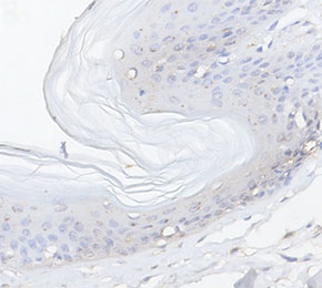 Immunohistochemical analysis of formalin fixed paraffin embedded human skin tissue with F4533 at 1:10 dilution.
キーポイント
この抗体には抗ラット二次抗体が必要です。
使用情報
| Dilution |
|---|
|
| Application |
|---|
| IHC, IF, FCM, ELISA |
| Source |
|---|
| Rat Monoclonal Antibody |
| Reactivity |
|---|
| Human, Mouse, Rat, Porcine, Equine |
| Storage Buffer |
|---|
| PBS, pH 7.2+50% Glycerol+0.05% BSA+0.01% NaN3 |
| Storage (from the date of receipt) |
|---|
| -20°C (avoid freeze-thaw cycles), 2 years |
| Predicted MW |
|---|
| 37 kDa |
| ポジティブコントロール | Human skin; Mouse epidermal sheet; K14E7 mouse epidermis; Porcine skin Langerhans cells; Cells derived from culture of porcine skin |
|---|---|
| ネガティブコントロール |
プロトコール
| IF |
|---|
Experimental Protocol:
Sample Preparation
1. Adherent Cells: Place a clean, sterile coverslip in a culture dish. Once the cells grow to near confluence as a monolayer, remove the coverslip for further use.
2. Suspension Cells: Seed the cells onto a clean, sterile slide coated with poly-L-lysine.
3. Frozen Sections: Allow the slide to thaw at room temperature. Wash it with pure water or PBS for 2 times, 3 minutes each time.
4. Paraffin Sections: Deparaffinization and rehydration. Wash the slide with pure water or PBS for 3 times, 3 minutes each time. Then perform antigen retrieval.
Fixation
1. Fix the cell coverslips/spots or tissue sections at room temperature using a fixative such as 4% paraformaldehyde (4% PFA) for 10-15 minutes.
2. Wash the sample with PBS for 3 times, 3 minutes each time.
Permeabilization
1.Add a detergent such as 0.1–0.3% Triton X-100 to the sample and incubate at room temperature for 10–20 minutes.
(Note: This step is only required for intracellular antigens. For antigens expressed on the cell membrane, this step is unnecessary.)
Wash the sample with PBS for 3 times, 3 minutes each time.
Blocking
Add blocking solution and incubate at room temperature for at least 1 hour. (Common blocking solutions include: serum from the same source as the secondary antibody, BSA, or goat serum.)
Note: Ensure the sample remains moist during and after the blocking step to prevent drying, which can lead to high background.
Immunofluorescence Staining (Day 1)
1. Remove the blocking solution and add the diluted primary antibody.
2. Incubate the sample in a humidified chamber at 4°C overnight.
Immunofluorescence Staining (Day 2)
1. Remove the primary antibody and wash with PBST for 3 times, 5 minutes each time.
2. Add the diluted fluorescent secondary antibody and incubate in the dark at 4°C for 1–2 hours.
3. Remove the secondary antibody and wash with PBST for 3 times, 5 minutes each time.
4. Add diluted DAPI and incubate at room temperature in the dark for 5–10 minutes.
5. Wash with PBST for 3 times, 5 minutes each time.
Mounting
1. Mount the sample with an anti-fade mounting medium.
2. Allow the slide to dry at room temperature overnight in the dark.
3. Store the slide in a slide storage box at 4°C, protected from light.
|
| IHC |
|---|
Experimental Protocol:
Deparaffinization/Rehydration
1. Deparaffinize/hydrate sections:
2. Incubate sections in three washes of xylene for 5 min each.
3. Incubate sections in two washes of 100% ethanol for 10 min each.
4. Incubate sections in two washes of 95% ethanol for 10 min each.
5. Wash sections two times in dH2O for 5 min each.
6.Antigen retrieval: For Citrate: Heat slides in a microwave submersed in 1X citrate unmasking solution until boiling is initiated; continue with 10 min at a sub-boiling temperature (95°-98°C). Cool slides on bench top for 30 min.
Staining
1. Wash sections in dH2O three times for 5 min each.
2. Incubate sections in 3% hydrogen peroxide for 10 min.
3. Wash sections in dH2O two times for 5 min each.
4. Wash sections in wash buffer for 5 min.
5. Block each section with 100–400 µl of blocking solution for 1 hr at room temperature.
6. Remove blocking solution and add 100–400 µl primary antibody diluent in to each section. Incubate overnight at 4°C.
7. Remove antibody solution and wash sections with wash buffer three times for 5 min each.
8. Cover section with 1–3 drops HRPas needed. Incubate in a humidified chamber for 30 min at room temperature.
9. Wash sections three times with wash buffer for 5 min each.
10. Add DAB Chromogen Concentrate to DAB Diluent and mix well before use.
11. Apply 100–400 µl DAB to each section and monitor closely. 1–10 min generally provides an acceptable staining intensity.
12. Immerse slides in dH2O.
13. If desired, counterstain sections with hematoxylin.
14. Wash sections in dH2O two times for 5 min each.
15. Dehydrate sections: Incubate sections in 95% ethanol two times for 10 sec each; Repeat in 100% ethanol, incubating sections two times for 10 sec each; Repeat in xylene, incubating sections two times for 10 sec each.
16. Mount sections with coverslips and mounting medium.
|
生物学的記述
| Specificity |
|---|
| Langerin/CD207 Antibody (Rat mAb) [F3M20] detects endogenous levels of total Langerin/CD207 protein. |
| タンパク質の局在 |
|---|
| 細胞内膜系 |
| Uniprot ID |
|---|
| Q9UJ71 |
| Clone |
|---|
| F3M20 |
| Synonym(s) |
|---|
| C-type lectin domain family 4 member K | C-type lectin domain family 4 | member K | CD207 | CD207 antigen | langerin | CD207 molecule | CD207 molecule | langerin | CLC4K | CLEC4K | Langerhans cell specific c-type lectin | Langerin |
| Background |
|---|
| Langerin (CD207) is a type II transmembrane C‑type lectin receptor selectively expressed by Langerhans cells and defined subsets of dendritic cells, where it functions as a sugar-binding endocytic receptor that links pathogen recognition at epithelial barriers to antigen processing and T‑cell activation. The protein comprises a short cytoplasmic tail, a single transmembrane segment, a coiled-coil neck that mediates homotrimer formation, and a luminal C‑type lectin carbohydrate recognition domain that binds mannose-, fucose-, and N‑acetylglucosamine-containing glycans and β‑glucans on the surfaces of viruses, fungi, and bacteria, and trimerization increases avidity and specificity for multivalent glycan ligands. Langerin localizes to the plasma membrane and to Birbeck granules, which are rod- or racket-shaped organelles formed by superimposed and zippered membranes whose biogenesis depends on Langerin expression and whose ultrastructure reflects Langerin-driven membrane rearrangements; mutational analysis shows that defined regions of Langerin are required for Birbeck granule formation, identifying this receptor as a key organizer of these LC-specific organelles. Ligand engagement induces rapid clathrin-independent internalization of Langerin–cargo complexes from the cell surface into Birbeck granules and associated endosomal structures, where captured antigens are processed and loaded onto MHC class II and, via cross-presentation, onto MHC class I molecules, and in vivo targeting of antigen to Langerin on dendritic cells results in efficient activation and proliferation of both CD4⁺ and CD8⁺ T cells with durable peptide–MHC display. This internalization and routing pathway allows Langerin to act as an antiviral and antifungal barrier at mucosal and cutaneous surfaces; HIV‑1, measles virus, and mycobacterial and fungal glycoconjugates are bound by Langerin and directed into Birbeck granules, where virions and microbial components are degraded and processed for antigen presentation rather than productively infecting Langerhans cells, thereby limiting pathogen transmission under conditions where Langerin expression and function are intact. Langerin is regulated by cytokines and tissue context, with TGF‑β1 and local epithelial factors promoting its expression during LC differentiation, and its expression is generally downregulated as Langerhans cells undergo terminal maturation and migration to lymph nodes, making CD207 a reliable marker of Langerhans cell identity and differentiation state in skin and mucosal epithelia as well as in specialized dendritic subsets in lymphoid and pulmonary tissues. Genetic variation in CD207 that reduces Langerin function or alters glycan binding compromises mannose recognition and is associated with increased susceptibility to cutaneous infection and changes in LC-mediated barrier immunity, and langerin expression is exploited diagnostically to distinguish Langerhans cell histiocytosis from non‑Langerhans histiocytic proliferations and to map Langerhans cell–rich inflammatory lesions in oral, skin, and pulmonary disease. |
| References |
|---|
技術サポート
ストックの作り方、阻害剤の保管方法、細胞実験や動物実験の際に注意すべき点など、製品を取扱う時に問い合わせが多かった質問に対しては取扱説明書でお答えしています。
他に質問がある場合は、お気軽にお問い合わせください。
* 必須

納期 国内在庫品:受注日の翌日(15時までの受注分) *北海道、九州、沖縄への配送は受注日より2日以上 を要する場合あり 海外在庫品:受注後1〜2週間