
- 阻害剤
- 研究分野別
- PI3K/Akt/mTOR
- Epigenetics
- Methylation
- Immunology & Inflammation
- Protein Tyrosine Kinase
- Angiogenesis
- Apoptosis
- Autophagy
- ER stress & UPR
- JAK/STAT
- MAPK
- Cytoskeletal Signaling
- Cell Cycle
- TGF-beta/Smad
- 化合物ライブラリー
- 抗体
- 新製品
- お問い合わせ

Mast Cell Chymase Antibody (Rabbit mAb) [C20M13]
CatNo: F8677
Application:
Reactivity:
-
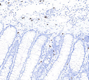 Immunohistochemical analysis of formalin fixed paraffin embedded human colon tissue with F8677 at 1:50 dilution.
当該製品は品切れ状态で、メールアドレスをご教示いただければ、お客様に返信いたします。
代表番号: 045-509-1970|電子メール:sales@selleck.co.jp
使用情報
| Dilution |
|---|
|
| Application |
|---|
| IHC |
| Source |
|---|
| Rabbit Monoclonal Antibody |
| Reactivity |
|---|
| Human |
| Storage Buffer |
|---|
| PBS, pH 7.2+50% Glycerol+0.05% BSA+0.01% NaN3 |
| Storage (from the date of receipt) |
|---|
| -20°C (avoid freeze-thaw cycles), 2 years |
| Predicted MW |
|---|
| 27 kDa |
| ポジティブコントロール | Human prostate adenocarcinoma; Human urothelial carcinoma; Human breast adenocarcinoma; Human endometrioid adenocarcinoma; Normal human colon; Normal human thymus; Normal human small intestine; Normal human esophagus; Human thymoma; Normal human uterus; Normal human lymph node; Normal human urothelial carcinoma; Normal human esophagus |
|---|---|
| ネガティブコントロール |
プロトコール
| IHC |
|---|
Experimental Protocol:
Deparaffinization/Rehydration
1. Deparaffinize/hydrate sections:
2. Incubate sections in three washes of xylene for 5 min each.
3. Incubate sections in two washes of 100% ethanol for 10 min each.
4. Incubate sections in two washes of 95% ethanol for 10 min each.
5. Wash sections two times in dH2O for 5 min each.
6.Antigen retrieval: For Citrate: Heat slides in a microwave submersed in 1X citrate unmasking solution until boiling is initiated; continue with 10 min at a sub-boiling temperature (95°-98°C). Cool slides on bench top for 30 min.
Staining
1. Wash sections in dH2O three times for 5 min each.
2. Incubate sections in 3% hydrogen peroxide for 10 min.
3. Wash sections in dH2O two times for 5 min each.
4. Wash sections in wash buffer for 5 min.
5. Block each section with 100–400 µl of blocking solution for 1 hr at room temperature.
6. Remove blocking solution and add 100–400 µl primary antibody diluent in to each section. Incubate overnight at 4°C.
7. Remove antibody solution and wash sections with wash buffer three times for 5 min each.
8. Cover section with 1–3 drops HRPas needed. Incubate in a humidified chamber for 30 min at room temperature.
9. Wash sections three times with wash buffer for 5 min each.
10. Add DAB Chromogen Concentrate to DAB Diluent and mix well before use.
11. Apply 100–400 µl DAB to each section and monitor closely. 1–10 min generally provides an acceptable staining intensity.
12. Immerse slides in dH2O.
13. If desired, counterstain sections with hematoxylin.
14. Wash sections in dH2O two times for 5 min each.
15. Dehydrate sections: Incubate sections in 95% ethanol two times for 10 sec each; Repeat in 100% ethanol, incubating sections two times for 10 sec each; Repeat in xylene, incubating sections two times for 10 sec each.
16. Mount sections with coverslips and mounting medium.
|
生物学的記述
| Specificity |
|---|
| Mast Cell Chymase Antibody (Rabbit mAb) [C20M13] detects endogenous levels of total Mast Cell Chymase protein. |
| Uniprot ID |
|---|
| P23946 |
| Clone |
|---|
| C20M13 |
| Synonym(s) |
|---|
| CMA1,CYH |
| Background |
|---|
| Mast cell chymase (chymase 1, CMA1) is a chymotrypsin-like serine protease stored in secretory granules of connective tissue–type mast cells and released upon activation, where it functions as a multifunctional effector enzyme that processes peptides, cytokines, and matrix components at sites of inflammation and tissue remodeling. The enzyme adopts the typical trypsin/chymotrypsin fold with a catalytic His–Asp–Ser triad in a substrate-binding cleft that prefers aromatic residues at the P1 position, and its heparin-dependent packing into mast cell granules positions chymase for rapid, localized proteolysis after degranulation. Angiotensin I represents a key physiological substrate, and human chymase efficiently converts angiotensin I to angiotensin II in the interstitial space, providing a major non–ACE pathway for angiotensin II generation in the heart and vasculature and contributing to local regulation of vascular tone, cardiac remodeling, and atherogenesis. Chymase also processes components of the extracellular matrix and matrix-regulating systems, including activation of matrix metalloproteinases and processing of type I procollagen, which link mast cell degranulation to collagen turnover, fibrotic matrix deposition, and structural changes in the vessel wall and parenchymal organs. Cytokine and growth factor bioavailability is strongly influenced by chymase, which can convert pro–IL‑1β to its active form, degrade IL‑4, and induce release of membrane-bound stem cell factor, thereby modulating local inflammatory cell recruitment, T helper cell polarization, and mast cell survival in a feedback-sensitive manner. Chymase functions as a chemoattractant for leukocytes and promotes migration of eosinophils and other inflammatory cells through protease-activated mechanisms and through remodeling of pericellular matrix, integrating mast cell activation with downstream recruitment and activation of effector leukocytes in allergic and chronic inflammatory settings. In airway disease, mast cell chymase contributes to airway smooth muscle and submucosal remodeling by degrading pericellular matrix, stimulating collagen fibril formation, and enhancing TGF‑β activation, aligning chymase activity with bronchial hyperresponsiveness, airway wall thickening, and features of asthma and chronic obstructive pulmonary disease. Cardiovascular pathologies, including hypertension, myocardial infarction, aneurysm formation, and valvular disease, are associated with increased mast cell chymase activity, where its angiotensin II–generating capacity, matrix-modifying actions, and pro-fibrotic signaling roles converge to drive adverse remodeling, neovascularization, and plaque instability. |
| References |
|---|
技術サポート
ストックの作り方、阻害剤の保管方法、細胞実験や動物実験の際に注意すべき点など、製品を取扱う時に問い合わせが多かった質問に対しては取扱説明書でお答えしています。
他に質問がある場合は、お気軽にお問い合わせください。
* 必須

納期 国内在庫品:受注日の翌日(15時までの受注分) *北海道、九州、沖縄への配送は受注日より2日以上 を要する場合あり 海外在庫品:受注後1〜2週間