
- 阻害剤
- 研究分野別
- PI3K/Akt/mTOR
- Epigenetics
- Methylation
- Immunology & Inflammation
- Protein Tyrosine Kinase
- Angiogenesis
- Apoptosis
- Autophagy
- ER stress & UPR
- JAK/STAT
- MAPK
- Cytoskeletal Signaling
- Cell Cycle
- TGF-beta/Smad
- 化合物ライブラリー
- 抗体
- 新製品
- お問い合わせ

MOB1 Antibody (Rabbit mAb) [P2B21]
CatNo: F0737
Application:
Reactivity:
-
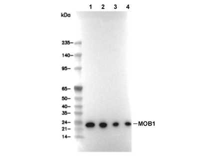 Lane 1: MOLT-4, Lane 2: Hela, Lane 3: COS-7, Lane 4: C6
当該製品は品切れ状态で、メールアドレスをご教示いただければ、お客様に返信いたします。
代表番号: 045-509-1970|電子メール:sales@selleck.co.jp
キーポイント
WB
転写条件(ウェット): 200 mA, 60 min。
転写条件(ウェット): 200 mA, 60 min。
使用情報
| Dilution |
|---|
|
| Application |
|---|
| WB, IP |
| Source |
|---|
| Rabbit Monoclonal Antibody |
| Reactivity |
|---|
| Human, Mouse, Rat, Hamster, Monkey |
| Storage Buffer |
|---|
| PBS, pH 7.2+50% Glycerol+0.05% BSA+0.01% NaN3 |
| Storage (from the date of receipt) |
|---|
| -20°C (avoid freeze-thaw cycles), 2 years |
| Predicted MW |
|---|
| 25 kDa |
| ポジティブコントロール | MOLT-4 cells; HeLa cells; PANC-1 cells; COS-7 cells; C2C12 cells; C6 cells; CHO cells |
|---|---|
| ネガティブコントロール |
プロトコール
| WB |
|---|
Experimental Protocol:
Sample preparation
1. Tissue: Lyse the tissue sample by adding an appropriate volume of ice-cold RIPA/NP-40 Lysis Buffer (containing Protease Inhibitor Cocktail),and homogenize the tissue at a low temperature or lyse it by sonication on ice, then incubate on ice for 30 minutes. 2. Adherent cell: Aspirate the culture medium and wash the cells with ice-cold PBS twice. Lyse the cells by adding an appropriate volume of RIPA/NP-40 Lysis Buffer (containing Protease Inhibitor Cocktail) , sonicate to lyse the cells, and incubate on ice for 30 minutes. 3. Suspension cell: Transfer the culture medium to a pre-cooled centrifuge tube. Centrifuge and aspirate the supernatant. Wash the cells with ice-cold PBS twice. Lyse the cells by adding an appropriate volume of RIPA/NP-40 Lysis Buffer (containing Protease Inhibitor Cocktail) , sonicate to lyse the cells, and incubate on ice for 30 minutes. 4. Place the lysate into a pre-cooled microcentrifuge tube. Centrifuge at 4°C for 15 min. Collect the supernatant;
5. Remove a small volume of lysate to determine the protein concentration;
6. Combine the lysate with protein loading buffer. Boil 20 µL sample under 95-100°C for 5 min. Centrifuge for 5 min after cool down on ice.
Electrophoretic separation
1. According to the concentration of extracted protein, load appropriate amount of protein sample and marker onto SDS-PAGE gels for electrophoresis. Recommended separating gel (lower gel) concentration: 10%. Reference Table for Selecting SDS-PAGE Separation Gel Concentrations 2. Power up 80V for 30 minutes. Then the power supply is adjusted (110 V~150 V), the Marker is observed, and the electrophoresis can be stopped when the indicator band of the predyed protein Marker where the protein is located is properly separated. (Note that the current should not be too large when electrophoresis, too large current (more than 150 mA) will cause the temperature to rise, affecting the result of running glue. If high currents cannot be avoided, an ice bath can be used to cool the bath.)
Transfer membrane
1. Take out the converter, soak the clip and consumables in the pre-cooled converter;
2. Activate PVDF membrane with methanol for 1 min and rinse with transfer buffer;
3. Install it in the order of "black edge of clip - sponge - filter paper - filter paper - glue -PVDF membrane - filter paper - filter paper - sponge - white edge of clip"; 4. The protein was electrotransferred to PVDF membrane. ( 0.45 µm PVDF membrane is recommended ) Reference Table for Selecting PVDF Membrane Pore Size Specifications Recommended conditions for wet transfer: 200 mA, 60 min. ( Note that the transfer conditions can be adjusted according to the protein size. For high-molecular-weight proteins, a higher current and longer transfer time are recommended. However, ensure that the transfer tank remains at a low temperature to prevent gel melting.)
Block
1. After electrotransfer, wash the film with TBST at room temperature for 5 minutes;
2. Incubate the film in the blocking solution for 1 hour at room temperature;
3. Wash the film with TBST for 3 times, 5 minutes each time.
Antibody incubation
1. Use primary antibody dilution buffer to prepare the primary antibody working liquid (recommended dilution ratio for primary antibody 1:1000), gently shake and incubate with the film at 4°C overnight; 2. Wash the film with TBST 3 times, 5 minutes each time;
3. Add the secondary antibody to the blocking solution and incubate with the film gently at room temperature for 1 hour;
4. After incubation, wash the film with TBST 3 times for 5 minutes each time.
Antibody staining
1. Add the prepared ECL luminescent substrate (or select other color developing substrate according to the second antibody) and mix evenly;
2. Incubate with the film for 1 minute, remove excess substrate (keep the film moist), wrap with plastic film, and expose in the imaging system. |
生物学的記述
| Specificity |
|---|
| MOB1 Antibody (Rabbit mAb) [P2B21] detects endogenous levels of total MOB1 protein. |
| タンパク質の局在 |
|---|
| 細胞質、細胞核 |
| Uniprot ID |
|---|
| Q7L9L4, Q9H8S9 |
| Clone |
|---|
| P2B21 |
| Synonym(s) |
|---|
| C2orf6 | FLJ10788 | FLJ11595 | MATS1 | MATS2 | MGC33910 | MOB kinase activator 1A | MOB kinase activator 1B | MOB1 | Mob1 alpha | Mob1 homolog 1A | Mob1 homolog 1B | MOB1 Mps One Binder homolog A | MOB1 Mps One Binder homolog B | MOB1 | Mps One Binder kinase activator-like 1A | MOB1 | Mps One Binder kinase activator-like 1A (yeast) | MOB1 | Mps One Binder kinase activator-like 1B | MOB1 | Mps One Binder kinase activator-like 1B (yeast) | Mob1A | Mob1B | MOB4A | MOB4B | MOBK1B | MOBKL1A | MOBKL1B | MOL1A | MOL1B | Mps one binder kinase activator-like 1A | Mps one binder kinase activator-like 1B | Protein Mob4A | Protein Mob4B |
| Background |
|---|
| MOB1, comprising the closely related human paralogs MOB1A and MOB1B, is a conserved kinase-activator protein that functions as a central adaptor in the Hippo tumor suppressor pathway and NDR/LATS kinase signaling, linking upstream MST/Hippo kinases to downstream regulators of cell proliferation, apoptosis, differentiation and cytokinesis. The protein adopts a compact MOB fold that binds the N‑terminal regulatory region of NDR and LATS family Ser/Thr kinases and contains several threonine residues that are phosphorylated by MST1/2, creating phospho‑dependent docking surfaces that strengthen MOB1–kinase interactions and stabilize active kinase conformations. Direct binding of human MOB1 to NDR kinases releases an autoinhibitory sequence in NDR, promotes phosphorylation of key regulatory sites in the activation segment and hydrophobic motif, and thereby converts NDR into a catalytically active state, establishing MOB1 as a bona fide kinase-activating subunit rather than a passive scaffold. In the canonical Hippo cascade, MOB1 associates with MST1/2 and coactivates LATS1/2, which phosphorylate and inhibit the transcriptional coactivators YAP and TAZ; loss of Mob1a/b in mouse keratinocytes reduces LATS activity, activates YAP1, and produces hyperproliferative, apoptosis-resistant keratinocyte progenitors with impaired contact inhibition and enhanced self‑renewal, demonstrating that MOB1 restrains tissue growth by enforcing Hippo-dependent control of YAP/TAZ. Mob1a/1b double-mutant mice exhibit embryonic lethality unless at least one wild-type allele is present, and conditional loss of the remaining allele drives trichilemmal carcinoma–like tumors with YAP1 activation, while some human trichilemmal carcinomas show MOB1A/B inactivation together with YAP1 activation, establishing MOB1 as a tumor suppressor whose dosage and integrity are critical for skin homeostasis and cancer susceptibility. In the lung, a MOB1–YAP1/TAZ–NKX2.1 axis controls bronchioalveolar epithelial differentiation, adhesion and tumor formation: inducible deletion of Mob1a/b in bronchioalveolar epithelium produces proliferative but poorly differentiated alveolar cells with reduced surfactant production and respiratory distress features, and reduces urethane-induced lung adenocarcinoma formation by depleting bronchioalveolar stem cells and lowering collagen XVII expression, indicating that MOB1 coordinates Hippo output and hemidesmosome components to maintain stem cell niches and tissue integrity. Beyond growth control, human MOB1A/B contribute to mitotic exit and cytokinesis by regulating midbody microtubule stability and centriole behavior; RNAi-mediated depletion of MOB1A/B causes abscission failure with hyperstable midbody microtubules, increased post-cytokinesis cell motility and prolonged centriole separation, whereas MOB1 overexpression prevents centrosome splitting, showing that MOB1 tunes microtubule dynamics at the intercellular bridge and centriole re-joining after telophase. At the signaling-network level, phospho‑MOB1 integrates inputs from MST/Hippo kinases and relays them to multiple NDR/LATS family members, allowing upstream stress, polarity and mechanical cues to be translated into coordinated changes in proliferation, apoptosis and cytoskeletal architecture. |
| References |
|---|
技術サポート
ストックの作り方、阻害剤の保管方法、細胞実験や動物実験の際に注意すべき点など、製品を取扱う時に問い合わせが多かった質問に対しては取扱説明書でお答えしています。
他に質問がある場合は、お気軽にお問い合わせください。
* 必須

納期 国内在庫品:受注日の翌日(15時までの受注分) *北海道、九州、沖縄への配送は受注日より2日以上 を要する場合あり 海外在庫品:受注後1〜2週間