
- 阻害剤
- 研究分野別
- PI3K/Akt/mTOR
- Epigenetics
- Methylation
- Immunology & Inflammation
- Protein Tyrosine Kinase
- Angiogenesis
- Apoptosis
- Autophagy
- ER stress & UPR
- JAK/STAT
- MAPK
- Cytoskeletal Signaling
- Cell Cycle
- TGF-beta/Smad
- 化合物ライブラリー
- 抗体
- 新製品
- お問い合わせ

NUP153 Antibody (Mouse mAb) [L18J13]
CatNo: F1570
Application:
Reactivity:
-
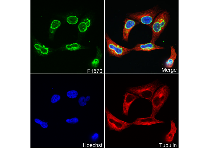 Immunofluorescent analysis of Hela cells using F1570 (green, 1:2000), Hoechst (blue) and tubulin (Red).
当該製品は品切れ状态で、メールアドレスをご教示いただければ、お客様に返信いたします。
代表番号: 045-509-1970|電子メール:sales@selleck.co.jp
使用情報
| Dilution |
|---|
|
| Application |
|---|
| IF |
| Source |
|---|
| Mouse Monoclonal Antibody |
| Reactivity |
|---|
| Mouse, Human |
| Storage Buffer |
|---|
| PBS, pH 7.2+50% Glycerol+0.05% BSA+0.01% NaN3 |
| Storage (from the date of receipt) |
|---|
| -20°C (avoid freeze-thaw cycles), 2 years |
| Predicted MW |
|---|
| 154 kDa |
| ポジティブコントロール | NIH3T3 cells; HeLa cells; U2OS cells; 3T3 cells |
|---|---|
| ネガティブコントロール |
プロトコール
| IF |
|---|
Experimental Protocol:
Sample Preparation
1. Adherent Cells: Place a clean, sterile coverslip in a culture dish. Once the cells grow to near confluence as a monolayer, remove the coverslip for further use.
2. Suspension Cells: Seed the cells onto a clean, sterile slide coated with poly-L-lysine.
3. Frozen Sections: Allow the slide to thaw at room temperature. Wash it with pure water or PBS for 2 times, 3 minutes each time.
4. Paraffin Sections: Deparaffinization and rehydration. Wash the slide with pure water or PBS for 3 times, 3 minutes each time. Then perform antigen retrieval.
Fixation
1. Fix the cell coverslips/spots or tissue sections at room temperature using a fixative such as 4% paraformaldehyde (4% PFA) for 10-15 minutes.
2. Wash the sample with PBS for 3 times, 3 minutes each time.
Permeabilization
1.Add a detergent such as 0.1–0.3% Triton X-100 to the sample and incubate at room temperature for 10–20 minutes.
(Note: This step is only required for intracellular antigens. For antigens expressed on the cell membrane, this step is unnecessary.)
Wash the sample with PBS for 3 times, 3 minutes each time.
Blocking
Add blocking solution and incubate at room temperature for at least 1 hour. (Common blocking solutions include: serum from the same source as the secondary antibody, BSA, or goat serum.)
Note: Ensure the sample remains moist during and after the blocking step to prevent drying, which can lead to high background.
Immunofluorescence Staining (Day 1)
1. Remove the blocking solution and add the diluted primary antibody.
2. Incubate the sample in a humidified chamber at 4°C overnight.
Immunofluorescence Staining (Day 2)
1. Remove the primary antibody and wash with PBST for 3 times, 5 minutes each time.
2. Add the diluted fluorescent secondary antibody and incubate in the dark at 4°C for 1–2 hours.
3. Remove the secondary antibody and wash with PBST for 3 times, 5 minutes each time.
4. Add diluted DAPI and incubate at room temperature in the dark for 5–10 minutes.
5. Wash with PBST for 3 times, 5 minutes each time.
Mounting
1. Mount the sample with an anti-fade mounting medium.
2. Allow the slide to dry at room temperature overnight in the dark.
3. Store the slide in a slide storage box at 4°C, protected from light.
|
生物学的記述
| Specificity |
|---|
| NUP153 Antibody (Mouse mAb) [L18J13] detects endogenous levels of total NUP153 protein. |
| Uniprot ID |
|---|
| P49790 |
| Clone |
|---|
| L18J13 |
| Synonym(s) |
|---|
| Nuclear pore complex protein Nup153 | 153 kDa nucleoporin | Nucleoporin Nup153 | NUP153 |
| Background |
|---|
| NUP153 is a vertebrate nucleoporin that belongs to the FG repeat–containing family of nuclear pore complex components and forms a major element of the nuclear basket, where it connects the central transport channel to peripheral nuclear structures and defines a key interface between the pore and the genome. The protein displays a modular organization with an N‑terminal region that anchors to the nuclear pore scaffold, a central Zn‑finger–rich segment, and a C‑terminal domain densely packed with FG repeats that interact with transport receptors and other nucleoporins to create a selective environment for macromolecular exchange across the nuclear envelope. Nup153 associates with the nuclear phase of the pore and participates in import and export of multiple cargo classes, including karyopherin‑dependent protein substrates and several classes of RNA, where it contributes to terminal docking steps and release events that shape the directionality and efficiency of nucleocytoplasmic transport. The protein also cooperates with Tpr and other basket nucleoporins to support retention and quality control of specific RNA species, integrating export capacity with surveillance functions that influence which transcripts reach the cytoplasm and at what rate. Nup153 establishes extensive contacts with chromatin and defines large nucleoporin‑associated domains that are enriched for euchromatic features, high RNA polymerase II occupancy, and transcriptionally active genes, linking nuclear pore components to genome‑wide transcriptional programs. These Nup153‑bound regions include clusters on the X chromosome in Drosophila that participate in dosage compensation, where Nup153 function is required for proper activity of the dosage compensation complex and for maintaining enhanced transcription output of X‑linked genes in males. Chromatin association by Nup153 spans genomic segments that range from tens to hundreds of kilobases, creating topological environments that support coordinated expression of gene neighborhoods rather than isolated promoters, and this organization contributes to the maintenance of broad transcriptionally active territories within the nucleus. Perturbation of Nup153 levels alters expression of a large fraction of the genome with pronounced down‑regulation within nucleoporin‑associated regions, demonstrating that this nucleoporin operates as a global transcriptional regulator in addition to its role in transport. |
| References |
|---|
技術サポート
ストックの作り方、阻害剤の保管方法、細胞実験や動物実験の際に注意すべき点など、製品を取扱う時に問い合わせが多かった質問に対しては取扱説明書でお答えしています。
他に質問がある場合は、お気軽にお問い合わせください。
* 必須

納期 国内在庫品:受注日の翌日(15時までの受注分) *北海道、九州、沖縄への配送は受注日より2日以上 を要する場合あり 海外在庫品:受注後1〜2週間