
- 阻害剤
- 研究分野別
- PI3K/Akt/mTOR
- Epigenetics
- Methylation
- Immunology & Inflammation
- Protein Tyrosine Kinase
- Angiogenesis
- Apoptosis
- Autophagy
- ER stress & UPR
- JAK/STAT
- MAPK
- Cytoskeletal Signaling
- Cell Cycle
- TGF-beta/Smad
- 化合物ライブラリー
- 抗体
- 新製品
- お問い合わせ

Orexin Antibody (Rabbit mAb) [F2B23]
CatNo: F6741
Application:
Reactivity:
-
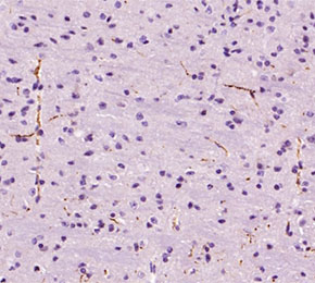 Immunohistochemical analysis of formalin fixed paraffin embedded mouse brain tissue with F6741 at 1:100 dilution.
当該製品は品切れ状态で、メールアドレスをご教示いただければ、お客様に返信いたします。
代表番号: 045-509-1970|電子メール:sales@selleck.co.jp
使用情報
| Dilution |
|---|
|
| Application |
|---|
| IHC, IF |
| Source |
|---|
| Rabbit Monoclonal Antibody |
| Reactivity |
|---|
| Human, Mouse, Rat |
| Storage Buffer |
|---|
| PBS, pH 7.2+50% Glycerol+0.05% BSA+0.01% NaN3 |
| Storage (from the date of receipt) |
|---|
| -20°C (avoid freeze-thaw cycles), 2 years |
| Predicted MW |
|---|
| 13 kDa |
| ポジティブコントロール | Human brain; Rat brain; Mouse hypothalamus |
|---|---|
| ネガティブコントロール |
プロトコール
| IF |
|---|
Experimental Protocol:
Sample Preparation
1. Adherent Cells: Place a clean, sterile coverslip in a culture dish. Once the cells grow to near confluence as a monolayer, remove the coverslip for further use.
2. Suspension Cells: Seed the cells onto a clean, sterile slide coated with poly-L-lysine.
3. Frozen Sections: Allow the slide to thaw at room temperature. Wash it with pure water or PBS for 2 times, 3 minutes each time.
4. Paraffin Sections: Deparaffinization and rehydration. Wash the slide with pure water or PBS for 3 times, 3 minutes each time. Then perform antigen retrieval.
Fixation
1. Fix the cell coverslips/spots or tissue sections at room temperature using a fixative such as 4% paraformaldehyde (4% PFA) for 10-15 minutes.
2. Wash the sample with PBS for 3 times, 3 minutes each time.
Permeabilization
1.Add a detergent such as 0.1–0.3% Triton X-100 to the sample and incubate at room temperature for 10–20 minutes.
(Note: This step is only required for intracellular antigens. For antigens expressed on the cell membrane, this step is unnecessary.)
Wash the sample with PBS for 3 times, 3 minutes each time.
Blocking
Add blocking solution and incubate at room temperature for at least 1 hour. (Common blocking solutions include: serum from the same source as the secondary antibody, BSA, or goat serum.)
Note: Ensure the sample remains moist during and after the blocking step to prevent drying, which can lead to high background.
Immunofluorescence Staining (Day 1)
1. Remove the blocking solution and add the diluted primary antibody.
2. Incubate the sample in a humidified chamber at 4°C overnight.
Immunofluorescence Staining (Day 2)
1. Remove the primary antibody and wash with PBST for 3 times, 5 minutes each time.
2. Add the diluted fluorescent secondary antibody and incubate in the dark at 4°C for 1–2 hours.
3. Remove the secondary antibody and wash with PBST for 3 times, 5 minutes each time.
4. Add diluted DAPI and incubate at room temperature in the dark for 5–10 minutes.
5. Wash with PBST for 3 times, 5 minutes each time.
Mounting
1. Mount the sample with an anti-fade mounting medium.
2. Allow the slide to dry at room temperature overnight in the dark.
3. Store the slide in a slide storage box at 4°C, protected from light.
|
| IHC |
|---|
Experimental Protocol:
Deparaffinization/Rehydration
1. Deparaffinize/hydrate sections:
2. Incubate sections in three washes of xylene for 5 min each.
3. Incubate sections in two washes of 100% ethanol for 10 min each.
4. Incubate sections in two washes of 95% ethanol for 10 min each.
5. Wash sections two times in dH2O for 5 min each.
6.Antigen retrieval: For Citrate: Heat slides in a microwave submersed in 1X citrate unmasking solution until boiling is initiated; continue with 10 min at a sub-boiling temperature (95°-98°C). Cool slides on bench top for 30 min.
Staining
1. Wash sections in dH2O three times for 5 min each.
2. Incubate sections in 3% hydrogen peroxide for 10 min.
3. Wash sections in dH2O two times for 5 min each.
4. Wash sections in wash buffer for 5 min.
5. Block each section with 100–400 µl of blocking solution for 1 hr at room temperature.
6. Remove blocking solution and add 100–400 µl primary antibody diluent in to each section. Incubate overnight at 4°C.
7. Remove antibody solution and wash sections with wash buffer three times for 5 min each.
8. Cover section with 1–3 drops HRPas needed. Incubate in a humidified chamber for 30 min at room temperature.
9. Wash sections three times with wash buffer for 5 min each.
10. Add DAB Chromogen Concentrate to DAB Diluent and mix well before use.
11. Apply 100–400 µl DAB to each section and monitor closely. 1–10 min generally provides an acceptable staining intensity.
12. Immerse slides in dH2O.
13. If desired, counterstain sections with hematoxylin.
14. Wash sections in dH2O two times for 5 min each.
15. Dehydrate sections: Incubate sections in 95% ethanol two times for 10 sec each; Repeat in 100% ethanol, incubating sections two times for 10 sec each; Repeat in xylene, incubating sections two times for 10 sec each.
16. Mount sections with coverslips and mounting medium.
|
生物学的記述
| Specificity |
|---|
| Orexin Antibody (Rabbit mAb) [F2B23] detects endogenous levels of total Orexin protein. |
| タンパク質の局在 |
|---|
| 細胞質小胞、小胞体、シナプス |
| Uniprot ID |
|---|
| O43612 |
| Clone |
|---|
| F2B23 |
| Synonym(s) |
|---|
| HCRT,PPOX |
| Background |
|---|
| Orexin (hypocretin) refers to a small family of hypothalamic neuropeptides produced from a single precursor (prepro-orexin) that is proteolytically processed into orexin-A and orexin-B, which act as high-affinity ligands for two rhodopsin-like G protein-coupled receptors, OX1R and OX2R, to coordinate arousal, energy balance, and motivated behaviors. Orexin neurons are clustered in the lateral and posterior hypothalamus and send dense projections throughout the brainstem and forebrain, including to monoaminergic and cholinergic arousal nuclei, the cortex, limbic regions, and autonomic centers, creating a widely distributed output network through which orexin peptides tonically stabilize wakefulness and phasic arousal by exciting noradrenergic, dopaminergic, histaminergic, and cholinergic systems. At the structural and receptor level, orexin-A and orexin-B share an N-terminal region and C-terminal amidation but differ in length and disulfide bonding, features that influence receptor selectivity, with OX1R showing preference for orexin-A and OX2R binding both peptides more equivalently; both receptors couple promiscuously to Gq, Gi/o, and Gs families in a cell-type–dependent manner, and robustly activate phospholipase cascades (PLC, PLA2, PLD) leading to IP3/Ca²⁺ mobilization, diacylglycerol and arachidonic acid production, and in some contexts diacylglycerol lipase–mediated synthesis of the endocannabinoid 2-arachidonoylglycerol that feeds back onto CB1 receptors. In central neurons, these signaling events converge on ion channels and exchangers, with inhibition of leak K⁺ currents and activation of the Na⁺/Ca²⁺ exchanger and nonselective cation channels producing strong depolarization and increased firing, a mechanism that underlies orexin-mediated excitation of wake-promoting nuclei, hypothalamic feeding circuits, and reward pathways. Orexin neurons integrate multiple afferent signals related to energy status (glucose, leptin, ghrelin), circadian phase, and stress, and adjust peptide output accordingly; elevated orexin signaling promotes wakefulness, physical activity, and food seeking, while loss of orexin or OX2R function causes narcolepsy with cataplexy in humans and animal models, directly linking orexin tone to sleep–wake stability. Beyond sleep and feeding, orexinergic projections to the hippocampus, amygdala, and prefrontal cortex modulate synaptic plasticity and memory processes, and receptor-specific pharmacology and mapping studies indicate that OX1R pathways have prominent roles in anxiety, pain modulation, and drug reward, whereas OX2R is particularly critical for non-REM and REM sleep regulation, making receptor subtype engagement an important determinant of functional output. Dysregulation of orexin signaling contributes to several disease contexts: deficiency or autoimmune loss of orexin neurons in narcolepsy; altered orexin and receptor expression patterns in obesity, addiction, depression, and neurodegenerative conditions; and aberrant orexin receptor activity in some cancers where orexin can induce caspase-dependent apoptosis. |
| References |
|---|
技術サポート
ストックの作り方、阻害剤の保管方法、細胞実験や動物実験の際に注意すべき点など、製品を取扱う時に問い合わせが多かった質問に対しては取扱説明書でお答えしています。
他に質問がある場合は、お気軽にお問い合わせください。
* 必須

納期 国内在庫品:受注日の翌日(15時までの受注分) *北海道、九州、沖縄への配送は受注日より2日以上 を要する場合あり 海外在庫品:受注後1〜2週間