
- 阻害剤
- 研究分野別
- PI3K/Akt/mTOR
- Epigenetics
- Methylation
- Immunology & Inflammation
- Protein Tyrosine Kinase
- Angiogenesis
- Apoptosis
- Autophagy
- ER stress & UPR
- JAK/STAT
- MAPK
- Cytoskeletal Signaling
- Cell Cycle
- TGF-beta/Smad
- 化合物ライブラリー
- 抗体
- 新製品
- お問い合わせ

Pericentrin Antibody (Rabbit mAb) [N1M2]
CatNo: F2239
Application:
Reactivity:
-
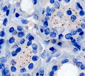 Immunohistochemical analysis of formalin fixed paraffin embedded human thyroid cancer tissue with F2239 at 1:2000 dilution.
当該製品は品切れ状态で、メールアドレスをご教示いただければ、お客様に返信いたします。
代表番号: 045-509-1970|電子メール:sales@selleck.co.jp
使用情報
| Dilution |
|---|
|
| Application |
|---|
| IHC, IF, FCM |
| Source |
|---|
| Rabbit Monoclonal Antibody |
| Reactivity |
|---|
| Mouse, Human |
| Storage Buffer |
|---|
| PBS, pH 7.2+50% Glycerol+0.05% BSA+0.01% NaN3 |
| Storage (from the date of receipt) |
|---|
| -20°C (avoid freeze-thaw cycles), 2 years |
| Predicted MW |
|---|
| 378 kDa |
| ポジティブコントロール | Human breast tissue; Human thyroid carcinoma tissue; Human pancreas tissue; NIH/3T3 cells; HeLa cells |
|---|---|
| ネガティブコントロール |
プロトコール
| IF |
|---|
Experimental Protocol:
Sample Preparation
1. Adherent Cells: Place a clean, sterile coverslip in a culture dish. Once the cells grow to near confluence as a monolayer, remove the coverslip for further use.
2. Suspension Cells: Seed the cells onto a clean, sterile slide coated with poly-L-lysine.
3. Frozen Sections: Allow the slide to thaw at room temperature. Wash it with pure water or PBS for 2 times, 3 minutes each time.
4. Paraffin Sections: Deparaffinization and rehydration. Wash the slide with pure water or PBS for 3 times, 3 minutes each time. Then perform antigen retrieval.
Fixation
1. Fix the cell coverslips/spots or tissue sections at room temperature using a fixative such as 4% paraformaldehyde (4% PFA) for 10-15 minutes.
2. Wash the sample with PBS for 3 times, 3 minutes each time.
Permeabilization
1.Add a detergent such as 0.1–0.3% Triton X-100 to the sample and incubate at room temperature for 10–20 minutes.
(Note: This step is only required for intracellular antigens. For antigens expressed on the cell membrane, this step is unnecessary.)
Wash the sample with PBS for 3 times, 3 minutes each time.
Blocking
Add blocking solution and incubate at room temperature for at least 1 hour. (Common blocking solutions include: serum from the same source as the secondary antibody, BSA, or goat serum.)
Note: Ensure the sample remains moist during and after the blocking step to prevent drying, which can lead to high background.
Immunofluorescence Staining (Day 1)
1. Remove the blocking solution and add the diluted primary antibody.
2. Incubate the sample in a humidified chamber at 4°C overnight.
Immunofluorescence Staining (Day 2)
1. Remove the primary antibody and wash with PBST for 3 times, 5 minutes each time.
2. Add the diluted fluorescent secondary antibody and incubate in the dark at 4°C for 1–2 hours.
3. Remove the secondary antibody and wash with PBST for 3 times, 5 minutes each time.
4. Add diluted DAPI and incubate at room temperature in the dark for 5–10 minutes.
5. Wash with PBST for 3 times, 5 minutes each time.
Mounting
1. Mount the sample with an anti-fade mounting medium.
2. Allow the slide to dry at room temperature overnight in the dark.
3. Store the slide in a slide storage box at 4°C, protected from light.
|
| IHC |
|---|
Experimental Protocol:
Deparaffinization/Rehydration
1. Deparaffinize/hydrate sections:
2. Incubate sections in three washes of xylene for 5 min each.
3. Incubate sections in two washes of 100% ethanol for 10 min each.
4. Incubate sections in two washes of 95% ethanol for 10 min each.
5. Wash sections two times in dH2O for 5 min each.
6.Antigen retrieval: For Citrate: Heat slides in a microwave submersed in 1X citrate unmasking solution until boiling is initiated; continue with 10 min at a sub-boiling temperature (95°-98°C). Cool slides on bench top for 30 min.
Staining
1. Wash sections in dH2O three times for 5 min each.
2. Incubate sections in 3% hydrogen peroxide for 10 min.
3. Wash sections in dH2O two times for 5 min each.
4. Wash sections in wash buffer for 5 min.
5. Block each section with 100–400 µl of blocking solution for 1 hr at room temperature.
6. Remove blocking solution and add 100–400 µl primary antibody diluent in to each section. Incubate overnight at 4°C.
7. Remove antibody solution and wash sections with wash buffer three times for 5 min each.
8. Cover section with 1–3 drops HRPas needed. Incubate in a humidified chamber for 30 min at room temperature.
9. Wash sections three times with wash buffer for 5 min each.
10. Add DAB Chromogen Concentrate to DAB Diluent and mix well before use.
11. Apply 100–400 µl DAB to each section and monitor closely. 1–10 min generally provides an acceptable staining intensity.
12. Immerse slides in dH2O.
13. If desired, counterstain sections with hematoxylin.
14. Wash sections in dH2O two times for 5 min each.
15. Dehydrate sections: Incubate sections in 95% ethanol two times for 10 sec each; Repeat in 100% ethanol, incubating sections two times for 10 sec each; Repeat in xylene, incubating sections two times for 10 sec each.
16. Mount sections with coverslips and mounting medium.
|
生物学的記述
| Specificity |
|---|
| Pericentrin Antibody (Rabbit mAb) [N1M2] detects endogenous levels of total Pericentrin protein. |
| タンパク質の局在 |
|---|
| 細胞質、細胞骨格、微小管 |
| Uniprot ID |
|---|
| O95613 |
| Clone |
|---|
| N1M2 |
| Synonym(s) |
|---|
| KIAA0402, PCNT2, PCNT, Pericentrin, Kendrin, Pericentrin-B |
| Background |
|---|
| Pericentrin is a large coiled-coil protein that forms an elongated scaffold within the pericentriolar material, the protein matrix surrounding the two centrioles that together constitute the centrosome, the principal microtubule organizing center of animal cells. As a structural scaffold, pericentrin anchors and recruits a defined set of pericentriolar material components, including CDK5RAP2, NEDD1, and the gamma-tubulin ring complex, positioning these microtubule-nucleating factors at the centrosome rather than allowing them to diffuse freely in the cytoplasm. Pericentrin-mediated anchoring of gamma-tubulin ring complexes at mitotic centrosomes is specifically required for proper spindle organization, and a pericentrin mutant with diminished binding to the gamma-tubulin complex components GCP2 and GCP3 shows significantly reduced centrosomal aster formation during mitosis, while interphase asters and Ran-mediated, centrosome-independent aster assembly remain largely unaffected, indicating that pericentrin's anchoring function is selectively required for mitotic, centrosome-dependent microtubule organization rather than for microtubule nucleation broadly. Loss of this anchoring mechanism triggers a checkpoint response that prevents mitotic entry and induces apoptotic cell death in a range of cell types, linking pericentrin-dependent centrosome integrity directly to cell cycle progression rather than solely to spindle geometry. Complete absence of pericentrin produces disorganized mitotic spindles and chromosome missegregation, and biallelic loss-of-function mutations in the PCNT gene cause microcephalic osteodysplastic primordial dwarfism type II, a condition marked by severe proportionate short stature and pronounced microcephaly despite near-normal intelligence in most affected individuals. Beyond spindle assembly, pericentrin contributes to centrosome-associated Chk1 recruitment, connecting centrosome integrity to G2/M checkpoint activation in the presence of DNA damage, and disruption of this checkpoint pathway represents one proposed mechanism by which pericentrin loss produces the growth failure characteristic of primordial dwarfism, alongside models implicating direct spindle orientation defects and randomized asymmetric versus symmetric stem cell divisions. Pericentrin overexpression, conversely, produces supernumerary centrosomes and multipolar spindles in fibroblasts, indicating that both loss and excess of pericentrin destabilize normal centrosome number and function. This dual sensitivity to pericentrin dosage, together with its established role as a scaffold-dependent checkpoint regulator rather than a passive structural marker, positions pericentrin as a defined target for researchers studying centrosome-linked growth disorders and the connection between centrosome integrity and genomic stability in cancer. |
| References |
|---|
技術サポート
ストックの作り方、阻害剤の保管方法、細胞実験や動物実験の際に注意すべき点など、製品を取扱う時に問い合わせが多かった質問に対しては取扱説明書でお答えしています。
他に質問がある場合は、お気軽にお問い合わせください。
* 必須

納期 国内在庫品:受注日の翌日(15時までの受注分) *北海道、九州、沖縄への配送は受注日より2日以上 を要する場合あり 海外在庫品:受注後1〜2週間