| PHLDA1 (pleckstrin homology‑like domain family A member 1, also known as TDAG51) is a proline‑ and histidine‑rich cytoplasmic protein that contains a split pleckstrin homology–like domain and belongs to the PHLDA family of phosphoinositide‑binding regulators of Akt signaling. The N‑ and C‑terminal segments of this PH‑like domain cooperate to bind phosphatidylinositol lipids at the inner leaflet of the plasma membrane, which positions PHLDA1 at membrane microdomains enriched in PI3K products and allows it to compete directly with Akt for access to these phosphoinositides. PHLDA1 acts as a negative regulator of the PI3K–Akt pathway; membrane recruitment through its PH‑like domain prevents efficient Akt docking and phosphorylation, reduces Akt activation, and attenuates downstream signaling to targets that control cell survival, proliferation, and metabolism, and PHLDA1 expression is induced by p53, linking this protein to a p53–PHLDA–Akt axis that connects stress responses to growth suppression. The protein is widely expressed in epithelial tissues, immune cells, endothelial cells, and neurons, and its abundance increases under diverse cellular stresses, including endoplasmic reticulum stress, oxidative stress, and loss of matrix attachment, where PHLDA1 participates in the regulation of apoptosis, anoikis, and other stress-related cell death programs through its influence on Akt and stress‑responsive transcription. PHLDA1 also modulates inflammatory signaling; in macrophages and related innate immune cells, it cooperates with the adaptor protein Tollip to suppress Toll‑like receptor 4–triggered production of proinflammatory cytokines, acting as a negative regulator of TLR4–NF‑κB activation and limiting excessive inflammatory responses during exposure to lipopolysaccharide. PHLDA1 decreased expression is associated with enhanced Akt activity, higher proliferative capacity, and poor prognosis in several carcinomas, consistent with a tumor‑suppressive function, while elevated PHLDA1 expression is reported in other malignancies where it aligns with oncogenic pathways and supports cell survival, indicating that the impact of PHLDA1 on tumor behavior depends on the surrounding signaling network. |

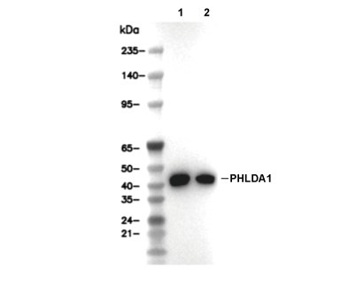 Lane 1: A375, Lane 2: BxPC3