
- 阻害剤
- 研究分野別
- PI3K/Akt/mTOR
- Epigenetics
- Methylation
- Immunology & Inflammation
- Protein Tyrosine Kinase
- Angiogenesis
- Apoptosis
- Autophagy
- ER stress & UPR
- JAK/STAT
- MAPK
- Cytoskeletal Signaling
- Cell Cycle
- TGF-beta/Smad
- 化合物ライブラリー
- 抗体
- 新製品
- お問い合わせ

Phospho-ABL1 (Tyr245) Antibody (Rabbit mAb) [F15J16]
CatNo: F0807
Application:
Reactivity:
-
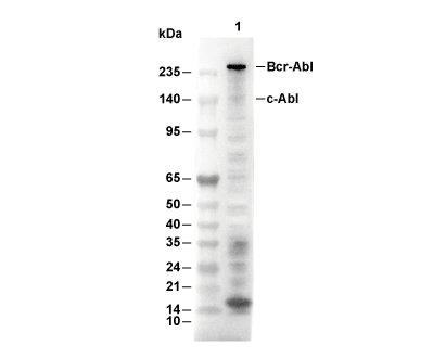 Lane 1: BV-173
当該製品は品切れ状态で、メールアドレスをご教示いただければ、お客様に返信いたします。
代表番号: 045-509-1970|電子メール:sales@selleck.co.jp
キーポイント
WB
SDS-PAGE の分離ゲルの推奨濃度:5%。
SDS-PAGE の分離ゲルの推奨濃度:5%。
使用情報
| Dilution |
|---|
|
| Application |
|---|
| WB |
| Source |
|---|
| Rabbit Monoclonal Antibody |
| Reactivity |
|---|
| Human |
| Storage Buffer |
|---|
| PBS, pH 7.2+50% Glycerol+0.05% BSA+0.01% NaN3 |
| Storage (from the date of receipt) |
|---|
| -20°C (avoid freeze-thaw cycles), 2 years |
| Predicted MW Observed MW |
|---|
| 123 kDa 135 kDa (c-Abl); 210 kDa (Bcr-Abl) |
| *なぜ予測分子量と実際の分子量が異なるのか? 下記の原因により、実際の分子量が予測と異なる:タンパク質の翻訳後修飾(リン酸化/糖鎖付加),スプライシングバリアント,イソフォーム,相対的な電荷,ポリマー。 |
| ポジティブコントロール | CML-T1 cells; BV173 cells |
|---|---|
| ネガティブコントロール |
プロトコール
| WB |
|---|
Experimental Protocol:
Sample preparation
1. Tissue: Lyse the tissue sample by adding an appropriate volume of ice-cold RIPA/NP-40 Lysis Buffer (containing Protease Inhibitor Cocktail, Phosphatase Inhibitor Cocktail),and homogenize the tissue at a low temperature or lyse it by sonication on ice, then incubate on ice for 30 minutes. 2. Adherent cell: Aspirate the culture medium and wash the cells with ice-cold PBS twice. Lyse the cells by adding an appropriate volume of RIPA/NP-40 Lysis Buffer (containing Protease Inhibitor Cocktail, Phosphatase Inhibitor Cocktail) , sonicate to lyse the cells, and incubate on ice for 30 minutes. 3. Suspension cell: Transfer the culture medium to a pre-cooled centrifuge tube. Centrifuge and aspirate the supernatant. Wash the cells with ice-cold PBS twice. Lyse the cells by adding an appropriate volume of RIPA/NP-40 Lysis Buffer (containing Protease Inhibitor Cocktail, Phosphatase Inhibitor Cocktail) , sonicate to lyse the cells, and incubate on ice for 30 minutes. 4. Place the lysate into a pre-cooled microcentrifuge tube. Centrifuge at 4°C for 15 min. Collect the supernatant;
5. Remove a small volume of lysate to determine the protein concentration;
6. Combine the lysate with protein loading buffer. Boil 20 µL sample under 95-100°C for 5 min. Centrifuge for 5 min after cool down on ice.
Electrophoretic separation
1. According to the concentration of extracted protein, load appropriate amount of protein sample and marker onto SDS-PAGE gels for electrophoresis. Recommended separating gel (lower gel) concentration: 5%. Reference Table for Selecting SDS-PAGE Separation Gel Concentrations 2. Power up 80V for 30 minutes. Then the power supply is adjusted (110 V~150 V), the Marker is observed, and the electrophoresis can be stopped when the indicator band of the predyed protein Marker where the protein is located is properly separated. (Note that the current should not be too large when electrophoresis, too large current (more than 150 mA) will cause the temperature to rise, affecting the result of running glue. If high currents cannot be avoided, an ice bath can be used to cool the bath.)
Transfer membrane
1. Take out the converter, soak the clip and consumables in the pre-cooled converter;
2. Activate PVDF membrane with methanol for 1 min and rinse with transfer buffer;
3. Install it in the order of "black edge of clip - sponge - filter paper - filter paper - glue -PVDF membrane - filter paper - filter paper - sponge - white edge of clip"; 4. The protein was electrotransferred to PVDF membrane. ( 0.45 µm PVDF membrane is recommended ) Reference Table for Selecting PVDF Membrane Pore Size Specifications Recommended conditions for wet transfer: 200 mA, 120 min. ( Note that the transfer conditions can be adjusted according to the protein size. For high-molecular-weight proteins, a higher current and longer transfer time are recommended. However, ensure that the transfer tank remains at a low temperature to prevent gel melting.)
Block
1. After electrotransfer, wash the film with TBST at room temperature for 5 minutes;
2. Incubate the film in the blocking solution ( recommending 5% BSA solution)
for 1 hour at room temperature;
3. Wash the film with TBST for 3 times, 5 minutes each time.
Antibody incubation
1. Use primary antibody dilution buffer to prepare the primary antibody working liquid (recommended dilution ratio for primary antibody 1:1000), gently shake and incubate with the film at 4°C overnight; 2. Wash the film with TBST 3 times, 5 minutes each time;
3. Add the secondary antibody to the blocking solution and incubate with the film gently at room temperature for 1 hour;
4. After incubation, wash the film with TBST 3 times for 5 minutes each time.
Antibody staining
1. Add the prepared ECL luminescent substrate (or select other color developing substrate according to the second antibody) and mix evenly;
2. Incubate with the film for 1 minute, remove excess substrate (keep the film moist), wrap with plastic film, and expose in the imaging system. |
生物学的記述
| Specificity |
|---|
| Phospho-ABL1 (Tyr245) Antibody (Rabbit mAb) [F15J16] detects endogenous levels of total ABL1 protein only when it is phosphorylated at Tyr245. |
| タンパク質の局在 |
|---|
| 細胞質、細胞骨格、細胞内膜系、ミトコンドリア、細胞核 |
| Uniprot ID |
|---|
| P00519 |
| Clone |
|---|
| F15J16 |
| Synonym(s) |
|---|
| Tyrosine-protein kinase ABL1, Abelson murine leukemia viral oncogene homolog 1, Abelson tyrosine-protein kinase 1, Proto-oncogene c-Abl, p150, ABL1, ABL, JTK7 |
| Background |
|---|
| * Phosphorylation of c-Abl at Tyr245 is a critical marker of activation of the non-receptor tyrosine kinase c-Abl, a multifunctional signaling protein of the Abl family that regulates cellular responses to DNA damage, oxidative stress, growth factors, adhesion signaling, and cytoskeletal dynamics in both the nucleus and cytoplasm. c-Abl contains an N-terminal cap region, SH3 and SH2 regulatory domains, a linker region harboring Tyr245, a catalytic kinase domain, and an activation loop containing Tyr412, which together maintain the kinase in an autoinhibited conformation under basal conditions. Phosphorylation at Tyr245 within the SH2-kinase linker disrupts inhibitory intramolecular interactions by repositioning the SH2 domain away from the catalytic cleft, thereby enhancing kinase activity and facilitating subsequent autophosphorylation at Tyr412 to stabilize the fully active conformation. Activated phospho-c-Abl phosphorylates multiple downstream substrates, including c-Jun to enhance AP-1–dependent transcription, CrkL to regulate Rac/Cdc42-mediated cytoskeletal remodeling, and Abi1 to control WAVE complex assembly and lamellipodia formation during cell migration. In DNA damage signaling, activated c-Abl interacts with ATM and promotes phosphorylation of pro-apoptotic factors such as p73, leading to induction of apoptotic genes including Puma, whereas during growth factor signaling such as PDGF stimulation, c-Abl cooperates with adaptor proteins like Nck and PI3K to activate MAPK/ERK pathways involved in proliferation and survival. Tyr245 phosphorylation is important in neuronal development, particularly in EphB receptor-mediated growth cone collapse and axon guidance, and also contributes to stress granule regulation through phosphorylation of RNA-binding proteins such as Sam68. Phosphorylation at Tyr245 induces a conformational shift that relieves steric inhibition of the ATP-binding pocket and activation loop, enabling efficient substrate access and kinase signaling. Dysregulated or constitutive Tyr245 phosphorylation is a hallmark of oncogenic BCR-ABL fusion proteins in leukemias, where persistent c-Abl activation drives Ras, STAT5, and PI3K signaling pathways that promote uncontrolled proliferation and myeloid transformation. |
| References |
|---|
技術サポート
ストックの作り方、阻害剤の保管方法、細胞実験や動物実験の際に注意すべき点など、製品を取扱う時に問い合わせが多かった質問に対しては取扱説明書でお答えしています。
他に質問がある場合は、お気軽にお問い合わせください。
* 必須

納期 国内在庫品:受注日の翌日(15時までの受注分) *北海道、九州、沖縄への配送は受注日より2日以上 を要する場合あり 海外在庫品:受注後1〜2週間