
- 阻害剤
- 研究分野別
- PI3K/Akt/mTOR
- Epigenetics
- Methylation
- Immunology & Inflammation
- Protein Tyrosine Kinase
- Angiogenesis
- Apoptosis
- Autophagy
- ER stress & UPR
- JAK/STAT
- MAPK
- Cytoskeletal Signaling
- Cell Cycle
- TGF-beta/Smad
- 化合物ライブラリー
- 抗体
- 新製品
- お問い合わせ
Phospho-AP2M1 (Thr156) Antibody [J20G9]
Catalog No.: F6265
Application:
Reactivity:
-
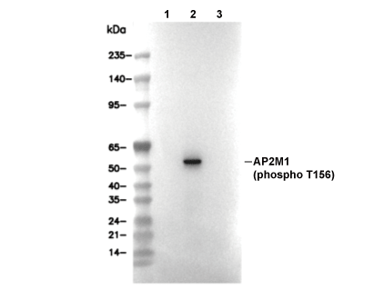 Lane 1: Hela (1% SDS Hot lysis), Lane 2: Hela (Calyculin A, 50 nM, 2 h; 1% SDS Hot lysis), Lane 3: Hela (Calyculin A, 50 nM, 2 h; 1% SDS Hot lysis; Alkaline Phosphatase, 1 h)
当該製品は品切れ状态で、メールアドレスをご教示いただければ、お客様に返信いたします。
代表番号: 045-509-1970|電子メール:sales@selleck.co.jp
キーポイント
WB
推奨WB希釈率: 1:10000
推奨WB希釈率: 1:10000
使用情報
| Dilution |
|---|
|
| Application |
|---|
| WB |
| Source |
|---|
| Rabbit Monoclonal Antibody |
| Reactivity |
|---|
| Mouse, Rat, Human |
| Storage Buffer |
|---|
| PBS, pH 7.2+50% Glycerol+0.05% BSA+0.01% NaN3 |
| Storage (from the date of receipt) |
|---|
| -20°C (avoid freeze-thaw cycles), 2 years |
| Predicted MW |
|---|
| 49 kDa |
| ポジティブコントロール | C6 cells (calyculin A, 100 ng/mL, 1 h; 1% SDS hot lysis method); HeLa cells (calyculin A, 50 nM, 2 h; 1% SDS hot lysis method); NIH/3T3 cells (calyculin A, 100 nM, 30 min) |
|---|---|
| ネガティブコントロール | C6 cells (calyculin A, 100 ng/mL, 1 h; RIPA lysis method); HeLa cells (calyculin A, 50 nM, 2 h; RIPA lysis method); C6 cells (1% SDS hot lysis method); HeLa cells (1% SDS hot lysis method); NIH/3T3 cells |
プロトコール
| WB |
|---|
Experimental Protocol:
Sample Preparation
1. Tissue samples: Disrupt the tissue, add an appropriate amount of preheated Hot 1% SDS Lysis Buffer (containing Protease Inhibitor Cocktail), and homogenize at 90 - 95℃. 2. Adherent cell samples: Aspirate the culture medium and wash the cells twice with ice-cold PBS. Add an appropriate amount of preheated Hot 1% SDS Lysis Buffer (containing Protease Inhibitor Cocktail), perform thermal lysis at 90 - 95℃ for 10 minutes, and repeatedly pipette to resuspend the cells during this period to ensure full contact between the cells and the hot lysis buffer. 3. Suspension cell: Transfer the culture medium to a pre-cooled centrifuge tube. Centrifuge and aspirate the supernatant. Wash the cells with ice-cold PBS twice.Add an appropriate amount of preheated Hot 1% SDS Lysis Buffer (containing Protease Inhibitor Cocktail), perform thermal lysis at 90 - 95℃ for 10 minutes, and repeatedly pipette to resuspend the cells during this period to ensure full contact between the cells and the hot lysis buffer. 4. Transfer the obtained homogenate/lysate to a centrifuge and centrifuge for 15 min, then collect the supernatant;
5. Take a small amount of the lysate to determine the protein concentration;
6. Add protein loading buffer, heat 20 μL of the sample at 95~100°C for 5 min, let it cool down on ice and then centrifuge for 5 min.
Electrophoretic separation
1. According to the concentration of extracted protein, load appropriate amount of protein sample and marker onto SDS-PAGE gels for electrophoresis. Recommended separating gel (lower gel) concentration: 10%. Reference Table for Selecting SDS-PAGE Separation Gel Concentrations 2. Power up 80V for 30 minutes. Then the power supply is adjusted (110 V~150 V), the Marker is observed, and the electrophoresis can be stopped when the indicator band of the predyed protein Marker where the protein is located is properly separated. (Note that the current should not be too large when electrophoresis, too large current (more than 150 mA) will cause the temperature to rise, affecting the result of running glue. If high currents cannot be avoided, an ice bath can be used to cool the bath.)
Transfer membrane
1. Take out the converter, soak the clip and consumables in the pre-cooled converter;
2. Activate PVDF membrane with methanol for 1 min and rinse with transfer buffer;
3. Install it in the order of "black edge of clip - sponge - filter paper - filter paper - glue -PVDF membrane - filter paper - filter paper - sponge - white edge of clip"; 4. The protein was electrotransferred to PVDF membrane. ( 0.45 µm PVDF membrane is recommended ) Reference Table for Selecting PVDF Membrane Pore Size Specifications Recommended conditions for wet transfer: 200 mA, 120 min. ( Note that the transfer conditions can be adjusted according to the protein size. For high-molecular-weight proteins, a higher current and longer transfer time are recommended. However, ensure that the transfer tank remains at a low temperature to prevent gel melting.)
Block
1. After electrotransfer, wash the film with TBST at room temperature for 5 minutes;
2. Incubate the film in the blocking solution ( recommending 5% BSA solution)
for 1 hour at room temperature;
3. Wash the film with TBST for 3 times, 5 minutes each time.
Antibody incubation
1. Use 5% skim milk powder to prepare the primary antibody working liquid (recommended dilution ratio for primary antibody 1:10000), gently shake and incubate with the film at 4°C overnight; 2. Wash the film with TBST 3 times, 5 minutes each time;
3. Add the secondary antibody to the blocking solution and incubate with the film gently at room temperature for 1 hour;
4. After incubation, wash the film with TBST 3 times for 5 minutes each time.
Antibody staining
1. Add the prepared ECL luminescent substrate (or select other color developing substrate according to the second antibody) and mix evenly;
2. Incubate with the film for 1 minute, remove excess substrate (keep the film moist), wrap with plastic film, and expose in the imaging system. |
生物学的記述
| Specificity |
|---|
| Phospho-AP2M1 (Thr156) Antibody [J20G9] detects endogenous levels of total AP2M1 protein only when it is phosphorylated at Thr156. |
| タンパク質の局在 |
|---|
| 細胞膜、被覆小窩、細胞内膜系 |
| Uniprot ID |
|---|
| Q96CW1 |
| Clone |
|---|
| J20G9 |
| Synonym(s) |
|---|
| AP-2 complex subunit mu; Adaptor-related protein complex 2 subunit mu; Clathrin coat-associated protein AP50; AP2M1 |
| Background |
|---|
| Phospho‑AP2M1 (Thr156) marks a key regulatory site on the μ2 subunit (AP2M1) of the AP‑2 clathrin adaptor complex, a heterotetrameric coat protein that couples cargo recognition to clathrin‑mediated endocytosis at the plasma membrane. AP2M1 lies at the core of the AP‑2 bent “V” architecture, where its C‑terminal domain forms the cargo‑binding interface that recognizes canonical YXXΦ and related endocytic sorting motifs in the cytoplasmic tails of transmembrane receptors, and its N‑terminal region participates in conformational transitions that couple cargo binding to clathrin‑lattice assembly. Phosphorylation of AP2M1 at Thr156 by the adaptor‑associated kinase AAK1 induces a conformational change in the AP‑2 complex that increases the accessibility and affinity of the μ2 cargo‑binding site for YXXΦ motifs, thereby enhancing the recruitment of cargo proteins such as the transferrin receptor into assembling clathrin‑coated pits and promoting efficient receptor‑mediated endocytosis. This phospho‑switch regulates the internalization and trafficking of signaling receptors, ion channels, and matrix‑remodeling enzymes, linking AP2M1 Thr156 phosphorylation to growth‑factor, neurotransmitter, and integrin‑linked pathways as well as to viral entry mechanisms that depend on clathrin‑mediated uptake. Dysregulation of AP2M1 Thr156 phosphorylation, driven either by AAK1 or by the Parkinson’s‑linked kinase LRRK2, alters endocytic capacity and trafficking of cargo receptors, producing endocytosis defects associated with neurodegeneration and perturbed receptor signaling. |
| References |
|---|
技術サポート
ストックの作り方、阻害剤の保管方法、細胞実験や動物実験の際に注意すべき点など、製品を取扱う時に問い合わせが多かった質問に対しては取扱説明書でお答えしています。
他に質問がある場合は、お気軽にお問い合わせください。
* 必須
納期 国内在庫品:受注日の翌日(15時までの受注分) *北海道、九州、沖縄への配送は受注日より2日以上 を要する場合あり 海外在庫品:受注後1〜2週間