
- 阻害剤
- 研究分野別
- PI3K/Akt/mTOR
- Epigenetics
- Methylation
- Immunology & Inflammation
- Protein Tyrosine Kinase
- Angiogenesis
- Apoptosis
- Autophagy
- ER stress & UPR
- JAK/STAT
- MAPK
- Cytoskeletal Signaling
- Cell Cycle
- TGF-beta/Smad
- 化合物ライブラリー
- 抗体
- 新製品
- お問い合わせ
Phospho-ULK1 (Ser555) Antibody [N1G15]
Catalog No.: F0305
Application:
Reactivity:
-
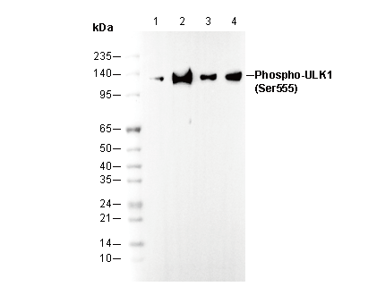 Lane 1: U-2 OS , Lane 2: U-2 OS (AMPK Activator, 50 μM, 1 hr) , Lane 3: MCF7 , Lane 4: MCF7 (oligomycin,0.5 μM,30 min)
カスタマーフィードバック(1)
キーポイント
WB
SDS-PAGE の分離ゲルの推奨濃度:5%。
このタンパク質の基底発現は低いため、事前に誘導刺激を行うことを推奨します。
SDS-PAGE の分離ゲルの推奨濃度:5%。
このタンパク質の基底発現は低いため、事前に誘導刺激を行うことを推奨します。
よくある問題とその対策
Fig. 1: WB (HepG2 Lysate) shows no bands (cells were not stimulated)
This antibody recognizes the Ser555-phosphorylated ULK1 protein. In some cell lines, the endogenous expression of Phospho-ULK1 (Ser555) is low without stimulation, leading to no visible bands. Therefore, it is recommended to stimulate the samples during preparation.
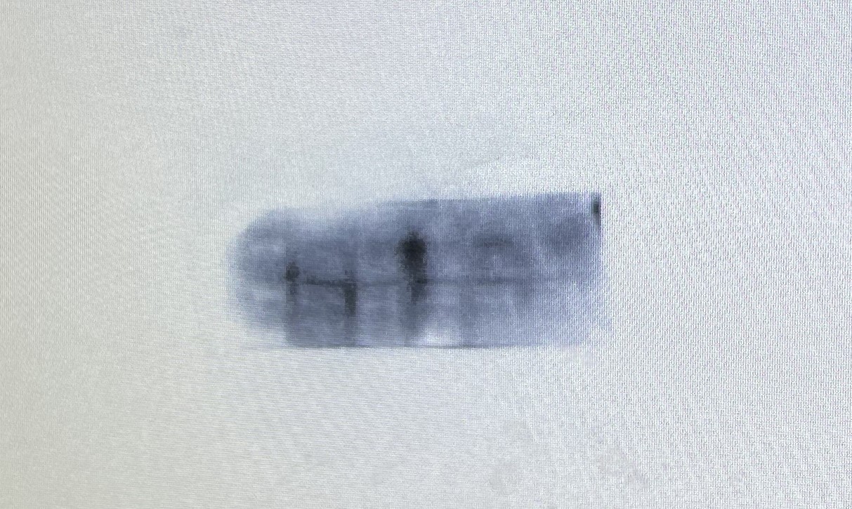
Fig. 2: WB (Human Pancreatic Cancer Cell Lysate) shows no bands (cells were not stimulated)
This antibody recognizes the Ser555-phosphorylated ULK1 protein, which is a key step in autophagy. In some cell lines, the endogenous expression of Phospho-ULK1 (Ser555) is low without stimulation. Therefore, it is recommended to stimulate the samples during preparation.
使用情報
| Dilution |
|---|
|
| Application |
|---|
| WB, IP |
| Source |
|---|
| Rabbit Monoclonal Antibody |
| Reactivity |
|---|
| Human, Mouse |
| Storage Buffer |
|---|
| PBS, pH 7.2+50% Glycerol+0.05% BSA+0.01% NaN₃ |
| Storage (from the date of receipt) |
|---|
| –20°C (avoid freeze-thaw cycles), 2 years |
| Predicted MW |
|---|
| 140-150 kDa |
| ポジティブコントロール | U-2 OS; MCF-7 (treated with CCCP, 100 μM, 2 h); C2C12 (treated with hydrogen peroxide, 10 mM, 5 min) |
|---|---|
| ネガティブコントロール | U-2 OS (ULK1 KO); MCF-7; C2C12 |
サンプル処理データの例
| サンプル | 処理状況 |
| U-2OS | AMPK Activator 991 (50 μM, 1 h) |
| MCF-7 | Oligomycin (0.5 μM, 30 min) |
| MCF-7 | Carbonyl cyanide 3-chlorophenylhydrazone (CCCP) (100 μM, 2 h) |
| クリックして、さらに多くのサンプルデータを表示 | |
*異なるヒト由来細胞や組織における発現量の予測については、以下をご参照ください: http://www.proteinatlas.org
プロトコール
| WB |
|---|
Experimental Protocol:
Sample preparation
1. Tissue: Lyse the tissue sample by adding an appropriate volume of ice-cold RIPA/NP-40 Lysis Buffer (containing Protease Inhibitor Cocktail, Phosphatase Inhibitor Cocktail),and homogenize the tissue at a low temperature or lyse it by sonication on ice, then incubate on ice for 30 minutes. 2. Adherent cell: Aspirate the culture medium and transfer the cells into an EP tube. Wash the cells with ice-cold PBS twice. Add an appropriate volume of RIPA/NP-40 Lysis Buffer (containing Protease Inhibitor Cocktail, Phosphatase Inhibitor Cocktail), sonicate to lyse the cells, and incubate on ice for 30 minutes. 3. Suspension cell: Transfer the culture medium to a pre-cooled centrifuge tube. Centrifuge and aspirate the supernatant. Wash the cells with ice-cold PBS twice.Add an appropriate volume of RIPA/NP-40 Lysis Buffer (containing Protease Inhibitor Cocktail, Phosphatase Inhibitor Cocktail), sonicate to lyse the cells, and incubate on ice for 30 minutes. 4. Place the lysate into a pre-cooled microcentrifuge tube. Centrifuge at 4°C for 15 min. Collect the supernatant;
5. Remove a small volume of lysate to determine the protein concentration;
6. Combine the lysate with protein loading buffer. Boil 20 µL sample under 95-100°C for 5 min. Centrifuge for 5 min after cool down on ice.
Electrophoretic separation
1. According to the concentration of extracted protein, load appropriate amount of protein sample and marker onto SDS-PAGE gels for electrophoresis. Recommended separating gel (lower gel) concentration: 5%. Reference Table for Selecting SDS-PAGE Separation Gel Concentrations 2. Power up 80V for 30 minutes. Then the power supply is adjusted (110 V~150 V), the Marker is observed, and the electrophoresis can be stopped when the indicator band of the predyed protein Marker where the protein is located is properly separated. (Note that the current should not be too large when electrophoresis, too large current (more than 150 mA) will cause the temperature to rise, affecting the result of running glue. If high currents cannot be avoided, an ice bath can be used to cool the bath.)
Transfer membrane
1. Take out the converter, soak the clip and consumables in the pre-cooled converter;
2. Activate PVDF membrane with methanol for 1 min and rinse with transfer buffer;
3. Install it in the order of "black edge of clip - sponge - filter paper - filter paper - glue -PVDF membrane - filter paper - filter paper - sponge - white edge of clip"; 4. The protein was electrotransferred to PVDF membrane. ( 0.45 µm PVDF membrane is recommended ) Reference Table for Selecting PVDF Membrane Pore Size Specifications Recommended conditions for wet transfer: 200 mA, 120 min. ( Note that the transfer conditions can be adjusted according to the protein size. For high-molecular-weight proteins, a higher current and longer transfer time are recommended. However, ensure that the transfer tank remains at a low temperature to prevent gel melting.)
Block
1. After electrotransfer, wash the film with TBST at room temperature for 5 minutes;
2. Incubate the film in the blocking solution ( recommending 5% BSA solution)
for 1 hour at room temperature;
3. Wash the film with TBST for 3 times, 5 minutes each time.
Antibody incubation
1. Use 5% skim milk powder to prepare the primary antibody working liquid (recommended dilution ratio for primary antibody 1:1000), gently shake and incubate with the film at 4°C overnight; 2. Wash the film with TBST 3 times, 5 minutes each time;
3. Add the secondary antibody to the blocking solution and incubate with the film gently at room temperature for 1 hour;
4. After incubation, wash the film with TBST 3 times for 5 minutes each time.
Antibody staining
36. Add the prepared ECL luminescent substrate (or select other color developing substrate according to the second antibody) and mix evenly;
2. Incubate with the film for 1 minute, remove excess substrate (keep the film moist), wrap with plastic film, and expose in the imaging system. |
生物学的記述
| Specificity |
|---|
Phospho-ULK1 (Ser555) Antibody [N1G15] recognizes endogenous levels of total Phospho-ULK1 (Ser555) protein. |
| タンパク質の局在 |
|---|
| 細胞質 |
| Uniprot ID |
|---|
| O75385 |
| Clone |
|---|
| N1G15 |
| Synonym(s) |
|---|
| Phospho-ULK1 (Ser555),ULK1 (phospho S555) |
| Background |
|---|
Phospho-ULK1 (Ser555) is a crucial phosphorylation site within the ULK1 protein complex, pivotal for regulating autophagy, a cellular process crucial for maintaining metabolic balance. AMP-activated protein kinase (AMPK) phosphorylates Ser555 in response to energy stress like glucose deprivation, enhancing ULK1 activity and promoting autophagy initiation. This phosphorylation also facilitates mitophagy, the selective degradation of damaged mitochondria, in response to cellular energy signals. Alongside Ser555, ULK1 undergoes various nutrient-sensitive phosphorylation events by AMPK and the mechanistic target of rapamycin complex 1 (MTORC1), highlighting the complex interplay between nutrient availability and autophagy regulation. |
| References |
|---|
技術サポート
ストックの作り方、阻害剤の保管方法、細胞実験や動物実験の際に注意すべき点など、製品を取扱う時に問い合わせが多かった質問に対しては取扱説明書でお答えしています。
他に質問がある場合は、お気軽にお問い合わせください。
* 必須
納期 国内在庫品:受注日の翌日(15時までの受注分) *北海道、九州、沖縄への配送は受注日より2日以上 を要する場合あり 海外在庫品:受注後1〜2週間