| Protein kinase C iota (PKCι), also designated PKCλ in mice, belongs to the atypical protein kinase C (aPKC) subfamily comprising PKCι and PKCζ, which exhibit calcium and diacylglycerol independence distinguishing them from conventional and novel PKC isoforms. PKCι contains an N-terminal regulatory domain featuring a single atypical cysteine-rich C1 domain lacking diacylglycerol binding capability, a Phox/Bem1 (PB1) domain mediating protein-protein interactions, and a pseudosubstrate motif, alongside a C-terminal catalytic serine/threonine kinase domain activated through phosphorylation at Thr500 in the activation loop, Thr641 via autophosphorylation, and Ser660 at the hydrophobic motif by PDK1 or related kinases. The protein exhibits ubiquitous tissue expression with particularly high levels in brain, lung, liver, and kidney, contrasting with the restricted expression pattern of PKCζ, and demonstrates 72% overall amino acid sequence identity to PKCζ despite exhibiting distinct, non-redundant cellular functions as evidenced by differential embryonic lethality upon genetic disruption. PKCι functions as a critical oncogene required for transformed growth, invasion, chemoresistance, and tumor cell survival across multiple human malignancies including non-small cell lung cancer, ovarian carcinoma, pancreatic adenocarcinoma, prostate cancer, colon carcinoma, and glioblastoma, with the PKCι gene on chromosome 3q26 representing a frequent target of tumor-specific gene amplification in approximately 36% of NSCLC cases and 70% of lung squamous cell carcinomas. PKCι drives oncogenic signaling through formation of a core complex with the scaffold protein Par6 and the small GTPase Rac1 or Cdc42, wherein the PB1 domain of PKCι heterodimerizes with the PB1 domain of Par6 while Par6's CRIB motif binds GTP-loaded Rac1 or Cdc42, coupling PKCι to polarity establishment, cytoskeletal reorganization, and transformed growth pathways. The PKCι-Par6-Rac1 polarity complex activates a Rac1→p21-activated kinase (PAK)→MEK→ERK signaling cascade essential for anchorage-independent transformed growth and tumorigenicity, with expression of kinase-deficient PKCι or constitutive disruption of the PKCι-Par6 interaction abolishing soft agar colony formation and tumor development without affecting adherent cell growth. PKCι promotes cell survival through at least three distinct mechanisms depending on cellular context—in chronic myelogenous leukemia cells, PKCι activates both canonical and non-canonical NF-κB signaling to induce IκB degradation and directly trans-activate nuclear NF-κB transcriptional activity conferring chemoresistance, in NSCLC cells Src-activated PKCι directly phosphorylates the pro-apoptotic protein Bad preventing its interaction with Bcl-XL to enhance survival, and in glioblastoma PKCι attenuates p38 MAPK signaling to reduce cisplatin-induced apoptosis. The protein functions downstream of oncogenic Ras, with Ras directly binding and activating PKCι, and PKCι proving essential for Ras-mediated transformation, invasion, and anchorage-independent growth in intestinal epithelial cells and critical for K-Ras-dependent colon carcinogenesis and lung tumor development. PKCι mediates cellular invasion through direct phosphorylation of μ- and m-calpains promoting wound healing and migration in NSCLC cells, and operates within a PKCβII→Ras→PKCι/Rac1→MEK signaling pathway driving invasive phenotypes in colon cancer cells. The PB1 domain contains a unique cysteine residue at position 69 (Cys69) located within the conserved OPCA motif at the PKCι-Par6 binding interface, where it interacts with Arg28 of Par6, and this cysteine serves as the molecular target for the selective PKCι inhibitor aurothiomalate (ATM) which forms a gold-cysteine adduct disrupting PKCι-Par6 and PKCι-p62 interactions without affecting other PB1 domain interactions. PKCι expression correlates with poor clinical outcome in NSCLC patients, with elevated PKCι conferring a 2.6-fold increased risk of cancer-related death and early-stage NSCLC patients with high PKCι exhibiting an 11-fold increased risk of relapse compared to those with low PKCι expression, positioning PKCι as both a prognostic biomarker and therapeutic target. Bcr-Abl transcriptionally activates PKCι through Ras/MEK-dependent induction of an Elk1 element within the proximal PKCι promoter, representing one mechanism of PKCι upregulation beyond gene amplification, although somatic mutations in PKCι appear absent or extremely rare based on sequencing analysis of all 18 exons in NSCLC cases. |

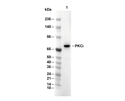 Lane 1: Mouse brain