
- 阻害剤
- 研究分野別
- PI3K/Akt/mTOR
- Epigenetics
- Methylation
- Immunology & Inflammation
- Protein Tyrosine Kinase
- Angiogenesis
- Apoptosis
- Autophagy
- ER stress & UPR
- JAK/STAT
- MAPK
- Cytoskeletal Signaling
- Cell Cycle
- TGF-beta/Smad
- 化合物ライブラリー
- 抗体
- 新製品
- お問い合わせ

PLK2 Antibody (Rabbit mAb) [M21F17]
CatNo: F6254
Application:
Reactivity:
-
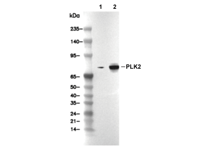 Lane 1: HCT 116, Lane 2: HCT 116 (Doxorubicin, 0.5 μM, 24 h)
当該製品は品切れ状态で、メールアドレスをご教示いただければ、お客様に返信いたします。
代表番号: 045-509-1970|電子メール:sales@selleck.co.jp
使用情報
| Dilution |
|---|
|
| Application |
|---|
| WB, IP |
| Source |
|---|
| Rabbit Monoclonal Antibody |
| Reactivity |
|---|
| Human |
| Storage Buffer |
|---|
| PBS, pH 7.2+50% Glycerol+0.05% BSA+0.01% NaN3 |
| Storage (from the date of receipt) |
|---|
| -20°C (avoid freeze-thaw cycles), 2 years |
| Predicted MW Observed MW |
|---|
| 78 kDa 70 kDa |
| *なぜ予測分子量と実際の分子量が異なるのか? 下記の原因により、実際の分子量が予測と異なる:タンパク質の翻訳後修飾(リン酸化/糖鎖付加),スプライシングバリアント,イソフォーム,相対的な電荷,ポリマー。 |
| ポジティブコントロール | ACHN cells; HCT 116 cells (Doxorubicin, 0.5 μM, 24 h) |
|---|---|
| ネガティブコントロール | HCT 116 cells |
プロトコール
| WB |
|---|
Experimental Protocol:
Sample preparation
1. Tissue: Lyse the tissue sample by adding an appropriate volume of ice-cold Tris-Triton Lysis Buffer (containing Protease Inhibitor Cocktail),and homogenize the tissue at a low temperature or lyse it by sonication on ice, then incubate on ice for 30 minutes. 2. Adherent cell: Aspirate the culture medium and wash the cells with ice-cold PBS twice. Lyse the cells by adding an appropriate volume of Tris-Triton Lysis Buffer (containing Protease Inhibitor Cocktail) , sonicate to lyse the cells, and incubate on ice for 30 minutes. 3. Suspension cell: Transfer the culture medium to a pre-cooled centrifuge tube. Centrifuge and aspirate the supernatant. Wash the cells with ice-cold PBS twice. Lyse the cells by adding an appropriate volume of Tris-Triton Lysis Buffer (containing Protease Inhibitor Cocktail) , sonicate to lyse the cells, and incubate on ice for 30 minutes. 4. Place the lysate into a pre-cooled microcentrifuge tube. Centrifuge at 4°C for 15 min. Collect the supernatant;
5. Remove a small volume of lysate to determine the protein concentration;
6. Combine the lysate with protein loading buffer. Boil 20 µL sample under 95-100°C for 5 min. Centrifuge for 5 min after cool down on ice.
Electrophoretic separation
1. According to the concentration of extracted protein, load appropriate amount of protein sample and marker onto SDS-PAGE gels for electrophoresis. Recommended separating gel (lower gel) concentration: 10%. Reference Table for Selecting SDS-PAGE Separation Gel Concentrations 2. Power up 80V for 30 minutes. Then the power supply is adjusted (110 V~150 V), the Marker is observed, and the electrophoresis can be stopped when the indicator band of the predyed protein Marker where the protein is located is properly separated. (Note that the current should not be too large when electrophoresis, too large current (more than 150 mA) will cause the temperature to rise, affecting the result of running glue. If high currents cannot be avoided, an ice bath can be used to cool the bath.)
Transfer membrane
1. Take out the converter, soak the clip and consumables in the pre-cooled converter;
2. Activate PVDF membrane with methanol for 1 min and rinse with transfer buffer;
3. Install it in the order of "black edge of clip - sponge - filter paper - filter paper - glue -PVDF membrane - filter paper - filter paper - sponge - white edge of clip"; 4. The protein was electrotransferred to PVDF membrane. ( 0.45 µm PVDF membrane is recommended ) Reference Table for Selecting PVDF Membrane Pore Size Specifications Recommended conditions for wet transfer: 200 mA, 120 min. ( Note that the transfer conditions can be adjusted according to the protein size. For high-molecular-weight proteins, a higher current and longer transfer time are recommended. However, ensure that the transfer tank remains at a low temperature to prevent gel melting.)
Block
1. After electrotransfer, wash the film with TBST at room temperature for 5 minutes;
2. Incubate the film in the blocking solution for 1 hour at room temperature;
3. Wash the film with TBST for 3 times, 5 minutes each time.
Antibody incubation
1. Use primary antibody dilution buffer to prepare the primary antibody working liquid (recommended dilution ratio for primary antibody 1:1000), gently shake and incubate with the film at 4°C overnight; 2. Wash the film with TBST 3 times, 5 minutes each time;
3. Add the secondary antibody to the blocking solution and incubate with the film gently at room temperature for 1 hour;
4. After incubation, wash the film with TBST 3 times for 5 minutes each time.
Antibody staining
1. Add the prepared ECL luminescent substrate (or select other color developing substrate according to the second antibody) and mix evenly;
2. Incubate with the film for 1 minute, remove excess substrate (keep the film moist), wrap with plastic film, and expose in the imaging system. |
生物学的記述
| Specificity |
|---|
| PLK2 Antibody (Rabbit mAb) [M21F17] detects endogenous levels of total PLK2 protein. |
| タンパク質の局在 |
|---|
| 細胞突起、細胞質、細胞骨格 |
| Uniprot ID |
|---|
| Q9NYY3 |
| Clone |
|---|
| M21F17 |
| Synonym(s) |
|---|
| hPlk2, hSNK, PLK-2, PLK2, polo like kinase 2, Polo-like kinase 2, polo-like kinase 2 (Drosophila), Serine/threonine-protein kinase PLK2, Serine/threonine-protein kinase SNK, Serum-inducible kinase, SNK |
| Background |
|---|
| Polo-like kinase 2 (PLK2), also known as serum- and glucocorticoid-inducible kinase SNK, is a serine/threonine kinase of the polo-like kinase family that links cell-cycle control and neuronal signaling by integrating p53-dependent checkpoint regulation, centriole duplication, and small GTPase-mediated synaptic plasticity. The protein shares the canonical organization of PLKs with an N-terminal catalytic kinase domain and C-terminal polo-box domains that recognize prephosphorylated motifs and localize PLK2 to defined subcellular structures, which permits context-specific phosphorylation of substrates involved in centrosome duplication, spindle checkpoint function, and Ras/Rap signaling complexes. During the cell cycle, PLK2 acts at the G1/S boundary and early mitotic events, where its kinase activity contributes to procentriole formation and accurate centriole replication, and its p53-inducible transcriptional control links centrosome duplication to DNA-damage and replication checkpoints, placing PLK2 in pathways that safeguard genomic integrity under proliferative and stress conditions. In neurons, PLK2 functions as a homeostatic suppressor of overexcitation and a key organizer of structural plasticity by coordinating the activity balance of Ras and Rap small GTPases at excitatory synapses; the kinase phosphorylates the Ras activator RASGRF1 and the Rap inhibitor SIPA1L1 to promote their degradation, while also phosphorylating the Rap activator RAPGEF2 and the Ras inhibitor SYNGAP1 to enhance their function, which shifts signaling away from Ras and toward Rap and thereby drives activity-dependent spine remodeling and adjusts synaptic strength. This bidirectional control of GEFs and GAP-like regulators situates PLK2 as an upstream timing and gain regulator for Ras–ERK and Rap-dependent cascades that influence AMPA receptor trafficking, dendritic spine density, and memory-related circuit refinement. PLK2 also modifies α-synuclein at regulatory sites that affect its turnover and aggregation, linking the kinase to proteostatic mechanisms that limit toxic protein accumulation and suggesting a functional interface between PLK2 activity, autophagic α-synuclein clearance, and neuronal resilience in synucleinopathies. PLK2 expression shows features of a stress-responsive kinase with context-dependent tumor suppressor properties; its p53-driven induction and roles in centriole duplication and checkpoint control connect PLK2 loss or silencing to centrosome abnormalities, cell-cycle deregulation, and chemoresistance, while retained or elevated PLK2 activity correlates with improved treatment response in specific cancer settings. |
| References |
|---|
技術サポート
ストックの作り方、阻害剤の保管方法、細胞実験や動物実験の際に注意すべき点など、製品を取扱う時に問い合わせが多かった質問に対しては取扱説明書でお答えしています。
他に質問がある場合は、お気軽にお問い合わせください。
* 必須

納期 国内在庫品:受注日の翌日(15時までの受注分) *北海道、九州、沖縄への配送は受注日より2日以上 を要する場合あり 海外在庫品:受注後1〜2週間