
- 阻害剤
- 研究分野別
- PI3K/Akt/mTOR
- Epigenetics
- Methylation
- Immunology & Inflammation
- Protein Tyrosine Kinase
- Angiogenesis
- Apoptosis
- Autophagy
- ER stress & UPR
- JAK/STAT
- MAPK
- Cytoskeletal Signaling
- Cell Cycle
- TGF-beta/Smad
- 化合物ライブラリー
- 抗体
- 新製品
- お問い合わせ
PLVAP/PV-1 Antibody [E23J22]
Catalog No.: F2799
Application:
Reactivity:
-
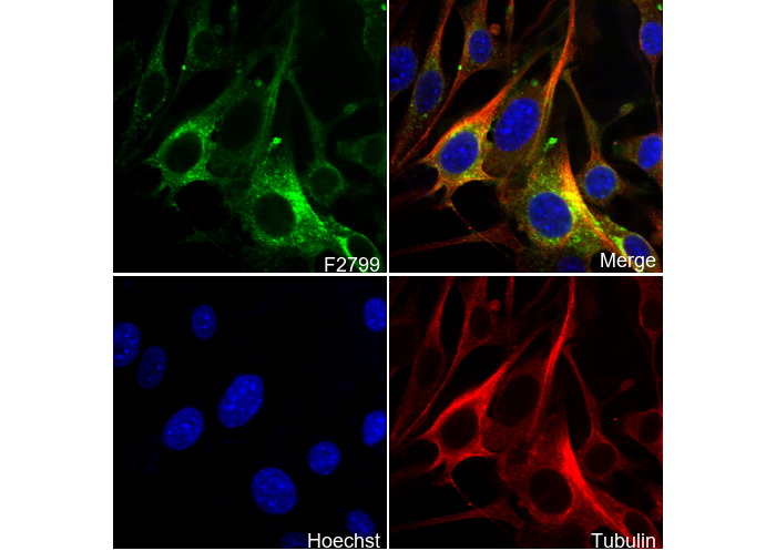 Immunofluorescent analysis of bEnd.3 cells using F2799 (green, 1:1000), Hoechst (blue) and tubulin (Red).
キーポイント
この抗体には抗ラット二次抗体が必要です。
使用情報
| Dilution |
|---|
|
| Application |
|---|
| IHC, IF, FCM |
| Source |
|---|
| Rat Monoclonal Antibody |
| Reactivity |
|---|
| Mouse |
| Storage Buffer |
|---|
| PBS, pH 7.2+50% Glycerol+0.05% BSA+0.01% NaN3 |
| Storage (from the date of receipt) |
|---|
| -20°C (avoid freeze-thaw cycles), 2 years |
| ポジティブコントロール | Mouse brain tissue; Mouse large intestine; bEND.3 cells |
|---|---|
| ネガティブコントロール |
プロトコール
| IF |
|---|
Experimental Protocol:
Sample Preparation
1. Adherent Cells: Place a clean, sterile coverslip in a culture dish. Once the cells grow to near confluence as a monolayer, remove the coverslip for further use.
2. Suspension Cells: Seed the cells onto a clean, sterile slide coated with poly-L-lysine.
3. Frozen Sections: Allow the slide to thaw at room temperature. Wash it with pure water or PBS for 2 times, 3 minutes each time.
4. Paraffin Sections: Deparaffinization and rehydration. Wash the slide with pure water or PBS for 3 times, 3 minutes each time. Then perform antigen retrieval.
Fixation
1. Fix the cell coverslips/spots or tissue sections at room temperature using a fixative such as 4% paraformaldehyde (4% PFA) for 10-15 minutes.
2. Wash the sample with PBS for 3 times, 3 minutes each time.
Permeabilization
1.Add a detergent such as 0.1–0.3% Triton X-100 to the sample and incubate at room temperature for 10–20 minutes.
(Note: This step is only required for intracellular antigens. For antigens expressed on the cell membrane, this step is unnecessary.)
Wash the sample with PBS for 3 times, 3 minutes each time.
Blocking
Add blocking solution and incubate at room temperature for at least 1 hour. (Common blocking solutions include: serum from the same source as the secondary antibody, BSA, or goat serum.)
Note: Ensure the sample remains moist during and after the blocking step to prevent drying, which can lead to high background.
Immunofluorescence Staining (Day 1)
1. Remove the blocking solution and add the diluted primary antibody.
2. Incubate the sample in a humidified chamber at 4°C overnight.
Immunofluorescence Staining (Day 2)
1. Remove the primary antibody and wash with PBST for 3 times, 5 minutes each time.
2. Add the diluted fluorescent secondary antibody and incubate in the dark at 4°C for 1–2 hours.
3. Remove the secondary antibody and wash with PBST for 3 times, 5 minutes each time.
4. Add diluted DAPI and incubate at room temperature in the dark for 5–10 minutes.
5. Wash with PBST for 3 times, 5 minutes each time.
Mounting
1. Mount the sample with an anti-fade mounting medium.
2. Allow the slide to dry at room temperature overnight in the dark.
3. Store the slide in a slide storage box at 4°C, protected from light.
|
| IHC |
|---|
Experimental Protocol:
Deparaffinization/Rehydration
1. Deparaffinize/hydrate sections:
2. Incubate sections in three washes of xylene for 5 min each.
3. Incubate sections in two washes of 100% ethanol for 10 min each.
4. Incubate sections in two washes of 95% ethanol for 10 min each.
5. Wash sections two times in dH2O for 5 min each.
6.Antigen retrieval: For Citrate: Heat slides in a microwave submersed in 1X citrate unmasking solution until boiling is initiated; continue with 10 min at a sub-boiling temperature (95°-98°C). Cool slides on bench top for 30 min.
Staining
1. Wash sections in dH2O three times for 5 min each.
2. Incubate sections in 3% hydrogen peroxide for 10 min.
3. Wash sections in dH2O two times for 5 min each.
4. Wash sections in wash buffer for 5 min.
5. Block each section with 100–400 µl of blocking solution for 1 hr at room temperature.
6. Remove blocking solution and add 100–400 µl primary antibody diluent in to each section. Incubate overnight at 4°C.
7. Remove antibody solution and wash sections with wash buffer three times for 5 min each.
8. Cover section with 1–3 drops HRPas needed. Incubate in a humidified chamber for 30 min at room temperature.
9. Wash sections three times with wash buffer for 5 min each.
10. Add DAB Chromogen Concentrate to DAB Diluent and mix well before use.
11. Apply 100–400 µl DAB to each section and monitor closely. 1–10 min generally provides an acceptable staining intensity.
12. Immerse slides in dH2O.
13. If desired, counterstain sections with hematoxylin.
14. Wash sections in dH2O two times for 5 min each.
15. Dehydrate sections: Incubate sections in 95% ethanol two times for 10 sec each; Repeat in 100% ethanol, incubating sections two times for 10 sec each; Repeat in xylene, incubating sections two times for 10 sec each.
16. Mount sections with coverslips and mounting medium.
|
生物学的記述
| Specificity |
|---|
| PLVAP/PV-1 Antibody [E23J22] detects endogenous levels of total PLVAP/PV-1 protein. |
| タンパク質の局在 |
|---|
| 細胞膜、細胞質、細胞内膜系 |
| Uniprot ID |
|---|
| Q91VC4 |
| Clone |
|---|
| E23J22 |
| Synonym(s) |
|---|
| Pv1; Plasmalemma vesicle-associated protein; MECA-32 antigen; Plasmalemma vesicle protein 1; PV-1 |
| Background |
|---|
| PLVAP (PV-1, plasmalemma vesicle-associated protein) is a type II transmembrane glycoprotein selectively expressed in fenestrated endothelia of kidney glomeruli, intestinal villi, liver sinusoids, and tumor neovasculature, but absent from the blood-brain barrier and continuous vessels, that organizes stomatal and fenestral diaphragms via its ~390-residue C-terminal extracellular coiled-coil domain. This domain forms rigid, parallel α-helical homodimers stabilized by five interchain disulfide bonds (Cys142, Cys153, Cys178, Cys199, Cys224), radiating as 8–10 spoke-like fibers spanning caveolar necks or fenestrae with precise 10–15 nm geometry. Functioning as a molecular sieve, PLVAP selectively restricts plasma protein transcytosis through caveolae and transendothelial channels, establishing a cutoff that allows passage of albumin and IgG while blocking larger complexes. VEGF/PI3K/p38MAPK signaling transcriptionally induces PLVAP during angiogenesis to widen caveolar openings and increase diaphragm density, which paradoxically enhances paracellular leakage via endothelial contractility. Genetic ablation of PLVAP results in 2–3-fold enlarged caveolae, loss of diaphragms, protein-losing enteropathy, hypoproteinemia, hypertriglyceridemia, and neonatal lethality from circulatory collapse, underscoring its essential role in maintaining basal permeability restriction. PLVAP is upregulated by hypoxia and VEGF, marking leaky tumor microvasculature that facilitates metastasis, and mediates vasogenic edema and leukocyte diapedesis in ischemia and sepsis. |
| References |
|---|
技術サポート
ストックの作り方、阻害剤の保管方法、細胞実験や動物実験の際に注意すべき点など、製品を取扱う時に問い合わせが多かった質問に対しては取扱説明書でお答えしています。
他に質問がある場合は、お気軽にお問い合わせください。
* 必須
納期 国内在庫品:受注日の翌日(15時までの受注分) *北海道、九州、沖縄への配送は受注日より2日以上 を要する場合あり 海外在庫品:受注後1〜2週間