
- 阻害剤
- 研究分野別
- PI3K/Akt/mTOR
- Epigenetics
- Methylation
- Immunology & Inflammation
- Protein Tyrosine Kinase
- Angiogenesis
- Apoptosis
- Autophagy
- ER stress & UPR
- JAK/STAT
- MAPK
- Cytoskeletal Signaling
- Cell Cycle
- TGF-beta/Smad
- 化合物ライブラリー
- 抗体
- 新製品
- お問い合わせ
Proglucagon Antibody [M22K5]
Catalog No.: F0810
Application:
Reactivity:
-
 Immunohistochemical analysis of formalin fixed paraffin embedded mouse pancreas tissue with F0810 at 1:400 dilution.
 -
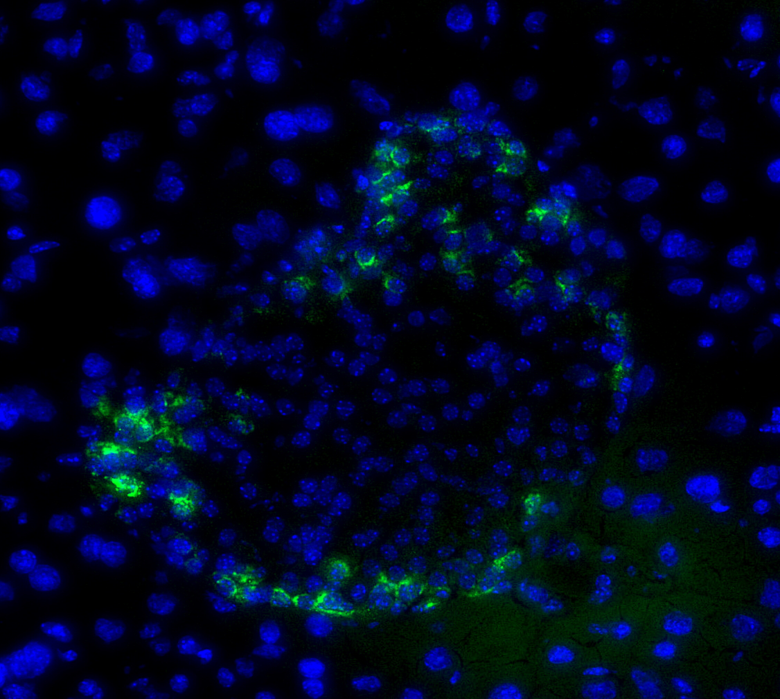 Immunofluorescent analysis of Mouse pancreatic tissue using F0810 (green, 1:400), Hoechst (blue) and tubulin (Red).
当該製品は品切れ状态で、メールアドレスをご教示いただければ、お客様に返信いたします。
代表番号: 045-509-1970|電子メール:sales@selleck.co.jp
使用情報
| Dilution |
|---|
|
| Application |
|---|
| IHC, IF |
| Source |
|---|
| Rabbit Monoclonal Antibody |
| Reactivity |
|---|
| Human, Mouse, Rat |
| Storage Buffer |
|---|
| PBS, pH 7.2+50% Glycerol+0.05% BSA+0.01% NaN3 |
| Storage (from the date of receipt) |
|---|
| -20°C (avoid freeze-thaw cycles), 2 years |
| ポジティブコントロール | Human pancreas; Rat pancreas; Mouse pancreas |
|---|---|
| ネガティブコントロール |
プロトコール
| IF |
|---|
Experimental Protocol:
Sample Preparation
1. Adherent Cells: Place a clean, sterile coverslip in a culture dish. Once the cells grow to near confluence as a monolayer, remove the coverslip for further use.
2. Suspension Cells: Seed the cells onto a clean, sterile slide coated with poly-L-lysine.
3. Frozen Sections: Allow the slide to thaw at room temperature. Wash it with pure water or PBS for 2 times, 3 minutes each time.
4. Paraffin Sections: Deparaffinization and rehydration. Wash the slide with pure water or PBS for 3 times, 3 minutes each time. Then perform antigen retrieval.
Fixation
1. Fix the cell coverslips/spots or tissue sections at room temperature using a fixative such as 4% paraformaldehyde (4% PFA) for 10-15 minutes.
2. Wash the sample with PBS for 3 times, 3 minutes each time.
Permeabilization
1.Add a detergent such as 0.1–0.3% Triton X-100 to the sample and incubate at room temperature for 10–20 minutes.
(Note: This step is only required for intracellular antigens. For antigens expressed on the cell membrane, this step is unnecessary.)
Wash the sample with PBS for 3 times, 3 minutes each time.
Blocking
Add blocking solution and incubate at room temperature for at least 1 hour. (Common blocking solutions include: serum from the same source as the secondary antibody, BSA, or goat serum.)
Note: Ensure the sample remains moist during and after the blocking step to prevent drying, which can lead to high background.
Immunofluorescence Staining (Day 1)
1. Remove the blocking solution and add the diluted primary antibody.
2. Incubate the sample in a humidified chamber at 4°C overnight.
Immunofluorescence Staining (Day 2)
1. Remove the primary antibody and wash with PBST for 3 times, 5 minutes each time.
2. Add the diluted fluorescent secondary antibody and incubate in the dark at 4°C for 1–2 hours.
3. Remove the secondary antibody and wash with PBST for 3 times, 5 minutes each time.
4. Add diluted DAPI and incubate at room temperature in the dark for 5–10 minutes.
5. Wash with PBST for 3 times, 5 minutes each time.
Mounting
1. Mount the sample with an anti-fade mounting medium.
2. Allow the slide to dry at room temperature overnight in the dark.
3. Store the slide in a slide storage box at 4°C, protected from light.
|
| IHC |
|---|
Experimental Protocol:
Deparaffinization/Rehydration
1. Deparaffinize/hydrate sections:
2. Incubate sections in three washes of xylene for 5 min each.
3. Incubate sections in two washes of 100% ethanol for 10 min each.
4. Incubate sections in two washes of 95% ethanol for 10 min each.
5. Wash sections two times in dH2O for 5 min each.
6.Antigen retrieval: For Citrate: Heat slides in a microwave submersed in 1X citrate unmasking solution until boiling is initiated; continue with 10 min at a sub-boiling temperature (95°-98°C). Cool slides on bench top for 30 min.
Staining
1. Wash sections in dH2O three times for 5 min each.
2. Incubate sections in 3% hydrogen peroxide for 10 min.
3. Wash sections in dH2O two times for 5 min each.
4. Wash sections in wash buffer for 5 min.
5. Block each section with 100–400 µl of blocking solution for 1 hr at room temperature.
6. Remove blocking solution and add 100–400 µl primary antibody diluent in to each section. Incubate overnight at 4°C.
7. Remove antibody solution and wash sections with wash buffer three times for 5 min each.
8. Cover section with 1–3 drops HRPas needed. Incubate in a humidified chamber for 30 min at room temperature.
9. Wash sections three times with wash buffer for 5 min each.
10. Add DAB Chromogen Concentrate to DAB Diluent and mix well before use.
11. Apply 100–400 µl DAB to each section and monitor closely. 1–10 min generally provides an acceptable staining intensity.
12. Immerse slides in dH2O.
13. If desired, counterstain sections with hematoxylin.
14. Wash sections in dH2O two times for 5 min each.
15. Dehydrate sections: Incubate sections in 95% ethanol two times for 10 sec each; Repeat in 100% ethanol, incubating sections two times for 10 sec each; Repeat in xylene, incubating sections two times for 10 sec each.
16. Mount sections with coverslips and mounting medium.
|
生物学的記述
| Specificity |
|---|
| Proglucagon Antibody [M22K5] detects endogenous levels of total Proglucagon protein. |
| タンパク質の局在 |
|---|
| 細胞外環境 |
| Uniprot ID |
|---|
| P01275 |
| Clone |
|---|
| M22K5 |
| Synonym(s) |
|---|
| Pro-glucagon;GCG |
| Background |
|---|
| Proglucagon, encoded by the Gcg gene, is a precursor polypeptide that serves as the source of multiple peptide hormones with important roles in metabolism and gut function. It is synthesized as a single chain protein of approximately 158–160 amino acids, with the glucagon sequence embedded internally and flanked by N and C terminal regions that harbor additional bioactive peptide domains. In pancreatic α cells, proglucagon undergoes proteolytic cleavage by prohormone convertase 2 (PC2) at paired basic residues to yield mature glucagon, a 29 amino acid peptide that activates the glucagon receptor on hepatocytes and stimulates glycogenolysis and gluconeogenesis through cAMP–PKA signaling. In intestinal L cells and certain brainstem neurons, the same precursor is processed by prohormone convertase 1/3 (PC1/3) to generate glucagon like peptide 1 (GLP 1), glucagon like peptide 2 (GLP 2), oxyntomodulin, and glicentin, each of which engages distinct receptors and downstream pathways. GLP 1 acts on the GLP 1 receptor in pancreatic β cells and the central nervous system to enhance glucose dependent insulin secretion, suppress glucagon release, delay gastric emptying, and promote satiety, whereas GLP 2 signals via the GLP 2 receptor on intestinal epithelial and subepithelial cells to support mucosal growth and barrier integrity. The tissue specific expression of PC2 and PC1/3 determines which peptide products dominate in the pancreas versus the gut, thereby shaping the balance between glucagon driven catabolism and GLP 1–mediated anabolic and gut protective effects. Dysregulated proglucagon processing or peptide secretion contributes to metabolic disease, as inappropriate glucagon release in α cells can exacerbate fasting and postprandial hyperglycemia, while reduced GLP 1 secretion or receptor responsiveness in the enteroinsular axis is associated with impaired glucose control and weight gain. |
| References |
|---|
技術サポート
ストックの作り方、阻害剤の保管方法、細胞実験や動物実験の際に注意すべき点など、製品を取扱う時に問い合わせが多かった質問に対しては取扱説明書でお答えしています。
他に質問がある場合は、お気軽にお問い合わせください。
* 必須
納期 国内在庫品:受注日の翌日(15時までの受注分) *北海道、九州、沖縄への配送は受注日より2日以上 を要する場合あり 海外在庫品:受注後1〜2週間