
- 阻害剤
- 研究分野別
- PI3K/Akt/mTOR
- Epigenetics
- Methylation
- Immunology & Inflammation
- Protein Tyrosine Kinase
- Angiogenesis
- Apoptosis
- Autophagy
- ER stress & UPR
- JAK/STAT
- MAPK
- Cytoskeletal Signaling
- Cell Cycle
- TGF-beta/Smad
- 化合物ライブラリー
- 抗体
- 新製品
- お問い合わせ
PSMD2 Antibody [B5P6]
Catalog No.: F6850
Application:
Reactivity:
-
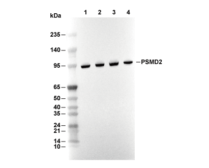 Lane 1: PC-3, Lane 2: OVCAR3, Lane 3: HCT 116, Lane 4: COS-7
当該製品は品切れ状态で、メールアドレスをご教示いただければ、お客様に返信いたします。
代表番号: 045-509-1970|電子メール:sales@selleck.co.jp
使用情報
| Dilution |
|---|
|
| Application |
|---|
| WB |
| Source |
|---|
| Rabbit Monoclonal Antibody |
| Reactivity |
|---|
| Human, Mouse, Rat, Monkey |
| Storage Buffer |
|---|
| PBS, pH 7.2+50% Glycerol+0.05% BSA+0.01% NaN3 |
| Storage (from the date of receipt) |
|---|
| -20°C (avoid freeze-thaw cycles), 2 years |
| Predicted MW |
|---|
| 97 kDa |
| ポジティブコントロール | PC-3 cells; RPMI 8226 cells; OVCAR3 cells; HCT 116 cells; LOX-IMVI cells; Hs578T cells; 786-0 cells; NCI-H23 cells; Hepa 1-6 cells; A-10 cells; COS-7 cells |
|---|---|
| ネガティブコントロール |
プロトコール
| WB |
|---|
Experimental Protocol:
Sample preparation
1. Tissue: Lyse the tissue sample by adding an appropriate volume of ice-cold RIPA/NP-40 Lysis Buffer (containing Protease Inhibitor Cocktail),and homogenize the tissue at a low temperature or lyse it by sonication on ice, then incubate on ice for 30 minutes. 2. Adherent cell: Aspirate the culture medium and wash the cells with ice-cold PBS twice. Lyse the cells by adding an appropriate volume of RIPA/NP-40 Lysis Buffer (containing Protease Inhibitor Cocktail), sonicate to lyse the cells, and incubate on ice for 30 minutes. 3. Suspension cell: Transfer the culture medium to a pre-cooled centrifuge tube. Centrifuge and aspirate the supernatant. Wash the cells with ice-cold PBS twice. Lyse the cells by adding an appropriate volume of RIPA/NP-40 Lysis Buffer (containing Protease Inhibitor Cocktail), sonicate to lyse the cells, and incubate on ice for 30 minutes. 4. Place the lysate into a pre-cooled microcentrifuge tube. Centrifuge at 4°C for 15 min. Collect the supernatant;
5. Remove a small volume of lysate to determine the protein concentration;
6. Combine the lysate with protein loading buffer. Boil 20 µL sample under 95-100°C for 5 min. Centrifuge for 5 min after cool down on ice.
Electrophoretic separation
1. According to the concentration of extracted protein, load appropriate amount of protein sample and marker onto SDS-PAGE gels for electrophoresis. Recommended separating gel (lower gel) concentration: 10%. Reference Table for Selecting SDS-PAGE Separation Gel Concentrations 2. Power up 80V for 30 minutes. Then the power supply is adjusted (110 V~150 V), the Marker is observed, and the electrophoresis can be stopped when the indicator band of the predyed protein Marker where the protein is located is properly separated. (Note that the current should not be too large when electrophoresis, too large current (more than 150 mA) will cause the temperature to rise, affecting the result of running glue. If high currents cannot be avoided, an ice bath can be used to cool the bath.)
Transfer membrane
1. Take out the converter, soak the clip and consumables in the pre-cooled converter;
2. Activate PVDF membrane with methanol for 1 min and rinse with transfer buffer;
3. Install it in the order of "black edge of clip - sponge - filter paper - filter paper - glue -PVDF membrane - filter paper - filter paper - sponge - white edge of clip"; 4. The protein was electrotransferred to PVDF membrane. ( 0.45 µm PVDF membrane is recommended ) Reference Table for Selecting PVDF Membrane Pore Size Specifications Recommended conditions for wet transfer: 200 mA, 120 min. ( Note that the transfer conditions can be adjusted according to the protein size. For high-molecular-weight proteins, a higher current and longer transfer time are recommended. However, ensure that the transfer tank remains at a low temperature to prevent gel melting.)
Block
1. After electrotransfer, wash the film with TBST at room temperature for 5 minutes;
2. Incubate the film in the blocking solution for 1 hour at room temperature;
3. Wash the film with TBST for 3 times, 5 minutes each time.
Antibody incubation
1. Use 5% skim milk powder to prepare the primary antibody working liquid (recommended dilution ratio for primary antibody 1:1000), gently shake and incubate with the film at 4°C overnight; 2. Wash the film with TBST 3 times, 5 minutes each time;
3. Add the secondary antibody to the blocking solution and incubate with the film gently at room temperature for 1 hour;
4. After incubation, wash the film with TBST 3 times for 5 minutes each time.
Antibody staining
1. Add the prepared ECL luminescent substrate (or select other color developing substrate according to the second antibody) and mix evenly;
2. Incubate with the film for 1 minute, remove excess substrate (keep the film moist), wrap with plastic film, and expose in the imaging system. |
生物学的記述
| Specificity |
|---|
| PSMD2 Antibody [B5P6] detects endogenous levels of total PSMD2 protein. |
| タンパク質の局在 |
|---|
| プロテアソーム |
| Uniprot ID |
|---|
| Q13200 |
| Clone |
|---|
| B5P6 |
| Synonym(s) |
|---|
| 26S proteasome non-ATPase regulatory subunit 2; 26S proteasome regulatory subunit RPN1; 26S proteasome subunit p97; Protein 55.11; PSMD2; TRAP2 |
| Background |
|---|
| PSMD2 (also known as Rpn1) is a non-ATPase subunit of the 19S regulatory particle (RP) lid within the 26S proteasome, playing a central scaffolding and ubiquitin receptor docking role essential for ATP- and ubiquitin-dependent degradation of misfolded proteins and regulatory factors, thereby ensuring cellular proteostasis. This large (~950 amino acids) protein contains PCI (proteasome-COP9 initiation) and 26S proteasome non-ATPase regulatory subunit domains, which together form a conformationally flexible scaffold at the periphery of the RP. PSMD2 features multiple PCI/MPN-binding surfaces that facilitate docking of ubiquitin shuttle proteins (such as Rad23 and Dsk2 via their UBL domains) and ubiquitin receptors (Rpn10 and Rpn13), effectively positioning ubiquitinated substrates for handoff to the ATPase base ring. PSMD2 coordinates the recognition of ubiquitin chains and their remodeling by lid-associated deubiquitinases (DUBs) like Rpn11, which, in concert with the AAA-ATPase ring (Rpt1-6), unfolds protein substrates and directs them through the gated 20S core β-subunits for proteolysis. As a result, the 26S proteasome processes approximately 70% of intracellular proteins, including key regulators of the cell cycle, apoptosis, and MHC class I antigen presentation. PSMD2 directly interacts with the intracellular domain of the TNF receptor, thereby linking proteasomal activity to the TNF/NF-κB signaling axis and contributing to immune regulation and inflammation. PSMD2 overexpression is associated with poor outcomes in lung adenocarcinoma and hepatocellular carcinoma, where it drives immune checkpoint modulation and resistance to therapy; conversely, PSMD2 inhibition selectively impairs proteasome chymotrypsin-like activity, inducing apoptosis in tumor cells while largely sparing healthy tissue. |
| References |
|---|
技術サポート
ストックの作り方、阻害剤の保管方法、細胞実験や動物実験の際に注意すべき点など、製品を取扱う時に問い合わせが多かった質問に対しては取扱説明書でお答えしています。
他に質問がある場合は、お気軽にお問い合わせください。
* 必須
納期 国内在庫品:受注日の翌日(15時までの受注分) *北海道、九州、沖縄への配送は受注日より2日以上 を要する場合あり 海外在庫品:受注後1〜2週間