
- 阻害剤
- 研究分野別
- PI3K/Akt/mTOR
- Epigenetics
- Methylation
- Immunology & Inflammation
- Protein Tyrosine Kinase
- Angiogenesis
- Apoptosis
- Autophagy
- ER stress & UPR
- JAK/STAT
- MAPK
- Cytoskeletal Signaling
- Cell Cycle
- TGF-beta/Smad
- 化合物ライブラリー
- 抗体
- 新製品
- お問い合わせ

REST Antibody (Mouse mAb) [B2F15]
Catalog No.: F4056
Application:
Reactivity:
-
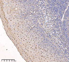 Immunohistochemical analysis of formalin fixed paraffin embedded human tonsils tissue with F4056 at 1:50 dilution.
当該製品は品切れ状态で、メールアドレスをご教示いただければ、お客様に返信いたします。
代表番号: 045-509-1970|電子メール:sales@selleck.co.jp
使用情報
| Dilution |
|---|
|
| Application |
|---|
| IHC, IF |
| Source |
|---|
| Mouse Monoclonal Antibody |
| Reactivity |
|---|
| Human |
| Storage Buffer |
|---|
| PBS, pH 7.2+50% Glycerol+0.05% BSA+0.01% NaN3 |
| Storage (from the date of receipt) |
|---|
| -20°C (avoid freeze-thaw cycles), 2 years |
| Predicted MW |
|---|
| 122 kDa |
| ポジティブコントロール | Human testis tissue; Human fallopian tube tissue; Human cortex tissue; Human tonsil tissue; Human liver tissue; Human kidney tissue; U-2 OS cells |
|---|---|
| ネガティブコントロール |
プロトコール
| IF |
|---|
Experimental Protocol:
Sample Preparation
1. Adherent Cells: Place a clean, sterile coverslip in a culture dish. Once the cells grow to near confluence as a monolayer, remove the coverslip for further use.
2. Suspension Cells: Seed the cells onto a clean, sterile slide coated with poly-L-lysine.
3. Frozen Sections: Allow the slide to thaw at room temperature. Wash it with pure water or PBS for 2 times, 3 minutes each time.
4. Paraffin Sections: Deparaffinization and rehydration. Wash the slide with pure water or PBS for 3 times, 3 minutes each time. Then perform antigen retrieval.
Fixation
1. Fix the cell coverslips/spots or tissue sections at room temperature using a fixative such as 4% paraformaldehyde (4% PFA) for 10-15 minutes.
2. Wash the sample with PBS for 3 times, 3 minutes each time.
Permeabilization
1.Add a detergent such as 0.1–0.3% Triton X-100 to the sample and incubate at room temperature for 10–20 minutes.
(Note: This step is only required for intracellular antigens. For antigens expressed on the cell membrane, this step is unnecessary.)
Wash the sample with PBS for 3 times, 3 minutes each time.
Blocking
Add blocking solution and incubate at room temperature for at least 1 hour. (Common blocking solutions include: serum from the same source as the secondary antibody, BSA, or goat serum.)
Note: Ensure the sample remains moist during and after the blocking step to prevent drying, which can lead to high background.
Immunofluorescence Staining (Day 1)
1. Remove the blocking solution and add the diluted primary antibody.
2. Incubate the sample in a humidified chamber at 4°C overnight.
Immunofluorescence Staining (Day 2)
1. Remove the primary antibody and wash with PBST for 3 times, 5 minutes each time.
2. Add the diluted fluorescent secondary antibody and incubate in the dark at 4°C for 1–2 hours.
3. Remove the secondary antibody and wash with PBST for 3 times, 5 minutes each time.
4. Add diluted DAPI and incubate at room temperature in the dark for 5–10 minutes.
5. Wash with PBST for 3 times, 5 minutes each time.
Mounting
1. Mount the sample with an anti-fade mounting medium.
2. Allow the slide to dry at room temperature overnight in the dark.
3. Store the slide in a slide storage box at 4°C, protected from light.
|
| IHC |
|---|
Experimental Protocol:
Deparaffinization/Rehydration
1. Deparaffinize/hydrate sections:
2. Incubate sections in three washes of xylene for 5 min each.
3. Incubate sections in two washes of 100% ethanol for 10 min each.
4. Incubate sections in two washes of 95% ethanol for 10 min each.
5. Wash sections two times in dH2O for 5 min each.
6.Antigen retrieval: For Citrate: Heat slides in a microwave submersed in 1X citrate unmasking solution until boiling is initiated; continue with 10 min at a sub-boiling temperature (95°-98°C). Cool slides on bench top for 30 min.
Staining
1. Wash sections in dH2O three times for 5 min each.
2. Incubate sections in 3% hydrogen peroxide for 10 min.
3. Wash sections in dH2O two times for 5 min each.
4. Wash sections in wash buffer for 5 min.
5. Block each section with 100–400 µl of blocking solution for 1 hr at room temperature.
6. Remove blocking solution and add 100–400 µl primary antibody diluent in to each section. Incubate overnight at 4°C.
7. Remove antibody solution and wash sections with wash buffer three times for 5 min each.
8. Cover section with 1–3 drops HRPas needed. Incubate in a humidified chamber for 30 min at room temperature.
9. Wash sections three times with wash buffer for 5 min each.
10. Add DAB Chromogen Concentrate to DAB Diluent and mix well before use.
11. Apply 100–400 µl DAB to each section and monitor closely. 1–10 min generally provides an acceptable staining intensity.
12. Immerse slides in dH2O.
13. If desired, counterstain sections with hematoxylin.
14. Wash sections in dH2O two times for 5 min each.
15. Dehydrate sections: Incubate sections in 95% ethanol two times for 10 sec each; Repeat in 100% ethanol, incubating sections two times for 10 sec each; Repeat in xylene, incubating sections two times for 10 sec each.
16. Mount sections with coverslips and mounting medium.
|
生物学的記述
| Specificity |
|---|
| REST Antibody (Mouse mAb) [B2F15] detects endogenous levels of total REST protein. |
| Uniprot ID |
|---|
| Q13127 |
| Clone |
|---|
| B2F15 |
| Synonym(s) |
|---|
| NRSF, XBR, REST, RE1-silencing transcription factor, Neural-restrictive silencer factor, X2 box repressor |
| Background |
|---|
| REST (RE1-silencing transcription factor), also known as NRSF, is a Kruppel-type zinc finger transcriptional repressor that binds the conserved 21–23 bp RE1/NRSE motif and functions as a master negative regulator of neuronal gene expression across development, adult brain plasticity, and neurodegeneration. The protein contains eight Cys2-His2 zinc fingers arranged in a central DNA-binding domain that contacts RE1 elements in promoter and enhancer regions, flanked by N‑ and C‑terminal repression domains that recruit distinct corepressor platforms, including mSin3A–HDAC1/2 and CoREST–HDAC1/2–LSD1–G9a complexes, enabling coordinated histone deacetylation, H3K4 demethylation, and H3K9 trimethylation to compact chromatin and silence target loci. REST occupies thousands of genomic sites in embryonic stem cells, neural progenitors, and differentiated neurons, directly repressing genes encoding ion channels, synaptic vesicle proteins, neurotransmitter receptors, transporters, and neuron-specific microRNAs, and thereby modulates synaptogenesis, axon guidance, membrane excitability, and structural plasticity while also targeting non-neuronal pathways such as JAK–STAT and HIF‑1 signaling in human brain. REST expression is high in pluripotent stem cells and neural progenitor cells, where it maintains a non-neuronal transcriptional program and prevents premature neurogenesis, and declines in a spatially and temporally controlled manner during terminal neuronal differentiation, allowing derepression of neuronal genes and acquisition of mature neuronal phenotypes; persistent REST in progenitors biases fate toward glial lineages and influences gliogenesis. REST abundance and activity are dynamically regulated by casein kinase 1–βTrCP-dependent ubiquitin–proteasome degradation, lysosomal–autophagic turnover, and nucleocytoplasmic trafficking, with context‑dependent nuclear accumulation in mature neurons in response to ischemia, seizures, and Huntington’s disease, or cytoplasmic sequestration by huntingtin–HAP1–RILP complexes under basal conditions. Within aging human cortex and hippocampus, REST reactivates at low levels in excitatory neurons, where it binds promoters of pro‑apoptotic, oxidative stress, and AD‑related genes (including those encoding BAX, PUMA, TNF‑pathway adaptors, γ‑secretase components, and CDK5 regulators) and represses their expression, conferring stress resistance, reduced vulnerability to amyloid and tau pathology, and association with preserved cognition and longevity even in the presence of Alzheimer-type lesions. Loss or reduction of REST in aging neurons correlates with shortened neuronal lifespan and transition from normal cognition to mild cognitive impairment and Alzheimer’s disease, and depletion of REST is also observed in frontotemporal dementia and dementia with Lewy bodies, supporting a general neuroprotective role of REST in late life. In contrast, excessive REST activation in post‑ischemic hippocampal neurons or in temporal lobe epilepsy drives epigenetic silencing of subsets of REST targets such as GluA2, GluN2B, KCC2, and HCN channels, promoting Ca²⁺‑permeable AMPA receptor expression, altered chloride homeostasis, and network hyperexcitability that contribute to excitotoxic neuronal death and epileptogenesis. REST-dependent remodeling also extends to noncoding RNA networks, repressing neuron‑specific microRNAs that themselves target multiple non-neuronal and apoptotic genes, thereby amplifying REST’s reach into metabolic, inflammatory, and survival pathways; transcriptomic analyses in Down syndrome brain, organoids, and neural cells identify REST-target gene sets enriched in JAK–STAT, HIF‑1, axon guidance, and fatty acid metabolism pathways, linking altered REST levels to neurogenic‑to‑gliogenic fate shifts, astrocyte activation, and metabolic imbalance. In neurodegenerative settings, REST integrates hypoxic and inflammatory cues with epigenetic output: in hypoxia and ischemia, nuclear REST is induced and binds RE1 sites at neuronal survival genes, while also engaging with TET3 and NSD3 to promote 5‑hydroxymethylcytosine and H3K36me3 at selected loci, indicating a dual capacity to repress or activate transcription depending on cofactor assembly. |
| References |
|---|
技術サポート
ストックの作り方、阻害剤の保管方法、細胞実験や動物実験の際に注意すべき点など、製品を取扱う時に問い合わせが多かった質問に対しては取扱説明書でお答えしています。
他に質問がある場合は、お気軽にお問い合わせください。
* 必須

納期 国内在庫品:受注日の翌日(15時までの受注分) *北海道、九州、沖縄への配送は受注日より2日以上 を要する場合あり 海外在庫品:受注後1〜2週間