
- 阻害剤
- 研究分野別
- PI3K/Akt/mTOR
- Epigenetics
- Methylation
- Immunology & Inflammation
- Protein Tyrosine Kinase
- Angiogenesis
- Apoptosis
- Autophagy
- ER stress & UPR
- JAK/STAT
- MAPK
- Cytoskeletal Signaling
- Cell Cycle
- TGF-beta/Smad
- 化合物ライブラリー
- 抗体
- 新製品
- お問い合わせ

VAP1 Antibody (Rat mAb) [G15D13]
CatNo: F3800
Application:
Reactivity:
-
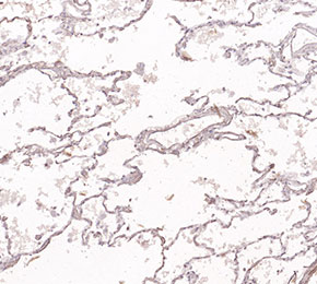 Immunohistochemical analysis of formalin fixed paraffin embedded mouse lung tissue with F3800 at 1:50 dilution.
当該製品は品切れ状态で、メールアドレスをご教示いただければ、お客様に返信いたします。
代表番号: 045-509-1970|電子メール:sales@selleck.co.jp
キーポイント
この抗体には抗ラット二次抗体が必要です。
使用情報
| Dilution |
|---|
|
| Application |
|---|
| IHC |
| Source |
|---|
| Rat Monoclonal Antibody |
| Reactivity |
|---|
| Mouse |
| Storage Buffer |
|---|
| PBS, pH 7.2+50% Glycerol+0.05% BSA+0.01% NaN3 |
| Storage (from the date of receipt) |
|---|
| -20°C (avoid freeze-thaw cycles), 2 years |
| Predicted MW |
|---|
| 85 kDa |
| ポジティブコントロール | Mouse muscle tissue |
|---|---|
| ネガティブコントロール |
プロトコール
| IF |
|---|
Experimental Protocol:
Sample Preparation
1. Adherent Cells: Place a clean, sterile coverslip in a culture dish. Once the cells grow to near confluence as a monolayer, remove the coverslip for further use.
2. Suspension Cells: Seed the cells onto a clean, sterile slide coated with poly-L-lysine.
3. Frozen Sections: Allow the slide to thaw at room temperature. Wash it with pure water or PBS for 2 times, 3 minutes each time.
4. Paraffin Sections: Deparaffinization and rehydration. Wash the slide with pure water or PBS for 3 times, 3 minutes each time. Then perform antigen retrieval.
Fixation
1. Fix the cell coverslips/spots or tissue sections at room temperature using a fixative such as 4% paraformaldehyde (4% PFA) for 10-15 minutes.
2. Wash the sample with PBS for 3 times, 3 minutes each time.
Permeabilization
1.Add a detergent such as 0.1–0.3% Triton X-100 to the sample and incubate at room temperature for 10–20 minutes.
(Note: This step is only required for intracellular antigens. For antigens expressed on the cell membrane, this step is unnecessary.)
Wash the sample with PBS for 3 times, 3 minutes each time.
Blocking
Add blocking solution and incubate at room temperature for at least 1 hour. (Common blocking solutions include: serum from the same source as the secondary antibody, BSA, or goat serum.)
Note: Ensure the sample remains moist during and after the blocking step to prevent drying, which can lead to high background.
Immunofluorescence Staining (Day 1)
1. Remove the blocking solution and add the diluted primary antibody.
2. Incubate the sample in a humidified chamber at 4°C overnight.
Immunofluorescence Staining (Day 2)
1. Remove the primary antibody and wash with PBST for 3 times, 5 minutes each time.
2. Add the diluted fluorescent secondary antibody and incubate in the dark at 4°C for 1–2 hours.
3. Remove the secondary antibody and wash with PBST for 3 times, 5 minutes each time.
4. Add diluted DAPI and incubate at room temperature in the dark for 5–10 minutes.
5. Wash with PBST for 3 times, 5 minutes each time.
Mounting
1. Mount the sample with an anti-fade mounting medium.
2. Allow the slide to dry at room temperature overnight in the dark.
3. Store the slide in a slide storage box at 4°C, protected from light.
|
生物学的記述
| Specificity |
|---|
VAP1 Antibody (Rat mAb) [G15D13] detects endogenous levels of total VAP1 protein. |
| タンパク質の局在 |
|---|
| 細胞膜、細胞内膜系 |
| Uniprot ID |
|---|
| Q16853 |
| Clone |
|---|
| G15D13 |
| Synonym(s) |
|---|
| Vap1, Amine oxidase [copper-containing] 3, Amine oxidase copper-containing 3, Copper amine oxidase, Semicarbazide-sensitive amine oxidase, Vascular adhesion protein 1, SSAO, VAP-1 |
| Background |
|---|
VAP-1, also designated semicarbazide-sensitive amine oxidase and encoded by the AOC3 gene, belongs to the copper-containing amine oxidase enzyme family and functions as a dual-purpose endothelial molecule, combining catalytic amine oxidase activity with a distinct role as an inducible leukocyte adhesion receptor. The enzyme catalyzes oxidative deamination of primary amines, including the endogenous substrates methylamine and aminoacetone, converting them to the corresponding aldehyde while releasing hydrogen peroxide and ammonia as reaction byproducts; catalysis depends on a copper ion together with a protein-derived topaquinone cofactor, distinguishing VAP-1 structurally and mechanistically from the flavin-dependent mitochondrial monoamine oxidases MAO-A and MAO-B despite the overlapping enzyme classification. Steady-state kinetic analysis using the model substrate benzylamine reveals a bell-shaped pH-dependence of the turnover rate, defined by two macroscopic ionizable groups within the enzyme-substrate complex, indicating that catalysis requires a specific protonation state maintained by residues positioned at the active site during amine turnover. VAP-1 exists in both a membrane-bound form on the surface of vascular endothelial and smooth muscle cells and a soluble circulating form generated through metalloproteinase-mediated proteolytic shedding of the membrane-anchored protein, and expression of membrane-bound VAP-1 is upregulated specifically at sites of vascular inflammation. Functioning as an adhesion molecule, VAP-1 mediates the slow-rolling and firm adhesion steps of the leukocyte adhesion cascade at high endothelial venules and inflamed blood vessels, supporting binding of lymphocytes, monocytes, and granulocytes to the endothelial surface as a prelude to transmigration into tissue, a role mechanistically distinct from its enzymatic amine-oxidizing function even though both activities reside on the same protein. In vascular smooth muscle cells, VAP-1 enzymatic activity toward aminoacetone and methylamine generates cytotoxic aldehyde products and contributes to oxidative stress within the vessel wall, extending VAP-1's pathological relevance beyond leukocyte recruitment into direct vascular tissue damage during atherosclerosis. VAP-1 is implicated in the chronic inflammatory component of atherosclerotic cardiovascular disease, including stroke and coronary artery disease, through its combined contribution to immune cell infiltration of the vessel wall and oxidative modification of vascular tissue, and genetic deletion of AOC3 in mice reduces leukocyte infiltration into adipose tissue, directly linking VAP-1 activity to tissue-specific immune cell recruitment in a model of metabolic and vascular inflammation. |
| References |
|---|
技術サポート
ストックの作り方、阻害剤の保管方法、細胞実験や動物実験の際に注意すべき点など、製品を取扱う時に問い合わせが多かった質問に対しては取扱説明書でお答えしています。
他に質問がある場合は、お気軽にお問い合わせください。
* 必須

納期 国内在庫品:受注日の翌日(15時までの受注分) *北海道、九州、沖縄への配送は受注日より2日以上 を要する場合あり 海外在庫品:受注後1〜2週間