
- 阻害剤
- 研究分野別
- PI3K/Akt/mTOR
- Epigenetics
- Methylation
- Immunology & Inflammation
- Protein Tyrosine Kinase
- Angiogenesis
- Apoptosis
- Autophagy
- ER stress & UPR
- JAK/STAT
- MAPK
- Cytoskeletal Signaling
- Cell Cycle
- TGF-beta/Smad
- 化合物ライブラリー
- 抗体
- 新製品
- お問い合わせ

VEGFA C-terminal Antibody (Rabbit mAb) [D1C9]
Catalog No.: F0064
Application:
Reactivity:
-
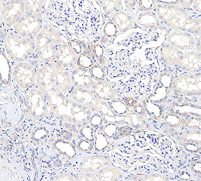 Immunohistochemical analysis of formalin fixed paraffin embedded human kidney tissue with F0064 at 1:100 dilution.
当該製品は品切れ状态で、メールアドレスをご教示いただければ、お客様に返信いたします。
代表番号: 045-509-1970|電子メール:sales@selleck.co.jp
使用情報
| Dilution |
|---|
|
| Application |
|---|
| IHC, IF, FCM |
| Source |
|---|
| Rabbit Monoclonal Antibody |
| Reactivity |
|---|
| Mouse, Human |
| Storage Buffer |
|---|
| PBS, pH 7.2+50% Glycerol+0.05% BSA+0.01% NaN3 |
| Storage (from the date of receipt) |
|---|
| -20°C (avoid freeze-thaw cycles), 2 years |
| Predicted MW |
|---|
| 44 kDa |
| ポジティブコントロール | Human kidney tissue; Mouse kidney tissue; NIH/3T3 cells |
|---|---|
| ネガティブコントロール |
プロトコール
| IF |
|---|
Experimental Protocol:
Sample Preparation
1. Adherent Cells: Place a clean, sterile coverslip in a culture dish. Once the cells grow to near confluence as a monolayer, remove the coverslip for further use.
2. Suspension Cells: Seed the cells onto a clean, sterile slide coated with poly-L-lysine.
3. Frozen Sections: Allow the slide to thaw at room temperature. Wash it with pure water or PBS for 2 times, 3 minutes each time.
4. Paraffin Sections: Deparaffinization and rehydration. Wash the slide with pure water or PBS for 3 times, 3 minutes each time. Then perform antigen retrieval.
Fixation
1. Fix the cell coverslips/spots or tissue sections at room temperature using a fixative such as 4% paraformaldehyde (4% PFA) for 10-15 minutes.
2. Wash the sample with PBS for 3 times, 3 minutes each time.
Permeabilization
1.Add a detergent such as 0.1–0.3% Triton X-100 to the sample and incubate at room temperature for 10–20 minutes.
(Note: This step is only required for intracellular antigens. For antigens expressed on the cell membrane, this step is unnecessary.)
Wash the sample with PBS for 3 times, 3 minutes each time.
Blocking
Add blocking solution and incubate at room temperature for at least 1 hour. (Common blocking solutions include: serum from the same source as the secondary antibody, BSA, or goat serum.)
Note: Ensure the sample remains moist during and after the blocking step to prevent drying, which can lead to high background.
Immunofluorescence Staining (Day 1)
1. Remove the blocking solution and add the diluted primary antibody.
2. Incubate the sample in a humidified chamber at 4°C overnight.
Immunofluorescence Staining (Day 2)
1. Remove the primary antibody and wash with PBST for 3 times, 5 minutes each time.
2. Add the diluted fluorescent secondary antibody and incubate in the dark at 4°C for 1–2 hours.
3. Remove the secondary antibody and wash with PBST for 3 times, 5 minutes each time.
4. Add diluted DAPI and incubate at room temperature in the dark for 5–10 minutes.
5. Wash with PBST for 3 times, 5 minutes each time.
Mounting
1. Mount the sample with an anti-fade mounting medium.
2. Allow the slide to dry at room temperature overnight in the dark.
3. Store the slide in a slide storage box at 4°C, protected from light.
|
| IHC |
|---|
Experimental Protocol:
Deparaffinization/Rehydration
1. Deparaffinize/hydrate sections:
2. Incubate sections in three washes of xylene for 5 min each.
3. Incubate sections in two washes of 100% ethanol for 10 min each.
4. Incubate sections in two washes of 95% ethanol for 10 min each.
5. Wash sections two times in dH2O for 5 min each.
6.Antigen retrieval: For Citrate: Heat slides in a microwave submersed in 1X citrate unmasking solution until boiling is initiated; continue with 10 min at a sub-boiling temperature (95°-98°C). Cool slides on bench top for 30 min.
Staining
1. Wash sections in dH2O three times for 5 min each.
2. Incubate sections in 3% hydrogen peroxide for 10 min.
3. Wash sections in dH2O two times for 5 min each.
4. Wash sections in wash buffer for 5 min.
5. Block each section with 100–400 µl of blocking solution for 1 hr at room temperature.
6. Remove blocking solution and add 100–400 µl primary antibody diluent in to each section. Incubate overnight at 4°C.
7. Remove antibody solution and wash sections with wash buffer three times for 5 min each.
8. Cover section with 1–3 drops HRPas needed. Incubate in a humidified chamber for 30 min at room temperature.
9. Wash sections three times with wash buffer for 5 min each.
10. Add DAB Chromogen Concentrate to DAB Diluent and mix well before use.
11. Apply 100–400 µl DAB to each section and monitor closely. 1–10 min generally provides an acceptable staining intensity.
12. Immerse slides in dH2O.
13. If desired, counterstain sections with hematoxylin.
14. Wash sections in dH2O two times for 5 min each.
15. Dehydrate sections: Incubate sections in 95% ethanol two times for 10 sec each; Repeat in 100% ethanol, incubating sections two times for 10 sec each; Repeat in xylene, incubating sections two times for 10 sec each.
16. Mount sections with coverslips and mounting medium.
|
生物学的記述
| Specificity |
|---|
| VEGFA C-terminal Antibody (Rabbit mAb) [D1C9] detects endogenous levels of total VEGFA protein. |
| Uniprot ID |
|---|
| P15692 |
| Clone |
|---|
| D1C9 |
| Synonym(s) |
|---|
| VEGF, VEGFA, L-VEGF, Vascular permeability factor, VPF |
| Background |
|---|
| The C-terminal domain of vascular endothelial growth factor A (VEGFA) determines isoform-specific properties through alternative splicing of exons 5, 7, 8a, and 8b, producing variants that retain similar affinity for VEGF receptor 2 (VEGFR2) but exhibit profound differences in binding to neuropilin-1 (NRP1) and heparan sulfate proteoglycans (HSPGs), which function as essential co-receptors modulating receptor presentation and signaling intensity. The C-terminal region encoded by exons 5 and 7 contains a heparin-binding domain (HBD) characterized by structural flexibility and two distinct basic amino acid clusters—residues R13, R14, K15 and residues K30, R35, R49—that cooperatively engage heparin and heparan sulfate through electrostatic interactions. This domain flexibility enables dynamic conformational changes essential for heparin binding, with C-terminal residues S34, C48, and D51 exhibiting millisecond-timescale motions that stabilize upon heparin octasaccharide engagement, facilitating receptor complex assembly and localization within the extracellular matrix. VEGFA isoforms containing the six amino acids encoded by exon 8a cooperate with domains from exons 5 or 7 to achieve efficient NRP1 and HSPG binding, whereas exon 8b-containing variants display divergent functional properties—VEGF165b binds VEGFR2 without triggering downstream phosphorylation cascades and functions as an angiogenesis inhibitor, while VEGF111b, the shortest exon 8b variant, paradoxically exhibits robust proangiogenic activity, illustrating that domain composition dictates functional outcomes through mechanisms beyond simple receptor affinity. The C-terminal domains regulate VEGFA bioavailability by controlling sequestration within the extracellular matrix through HSPG interactions and modulate receptor activation kinetics by recruiting NRP1, which enhances VEGFR2 signaling and promotes endothelial cell migration, proliferation, and survival through activation of PI3K/AKT and MAPK/ERK pathways. NRP1 binding initiated by the C-terminal domain extends VEGFA function beyond endothelial cells, mediating axon guidance and neuronal migration through distinct signaling networks involving semaphorin pathway components. The structural flexibility inherent to the C-terminal HBD permits interactions with diverse binding partners while maintaining specificity, enabling VEGFA to coordinate multiple cellular responses, including vascular permeability regulation, basement membrane remodeling, and integration with hypoxia-inducible factor signaling networks. Alternative splicing generating distinct C-terminal architectures creates a repertoire of VEGFA isoforms with graded angiogenic potency—matrix-bound longer isoforms establish localized signaling gradients directing vessel sprouting, while freely diffusible shorter variants promote distal endothelial cell activation, collectively orchestrating vessel patterning and maturation. Dysregulation of C-terminal domain expression patterns contributes to pathological angiogenesis, with tumor cells selectively producing exon 8a isoforms to maximize NRP1-mediated signaling and vascular permeability, facilitating nutrient delivery and metastatic dissemination, while therapeutic strategies targeting C-terminal domain functions through anti-VEGF antibodies or soluble decoy receptors disrupt both VEGFR2 and NRP1 engagement. |
| References |
|---|
技術サポート
ストックの作り方、阻害剤の保管方法、細胞実験や動物実験の際に注意すべき点など、製品を取扱う時に問い合わせが多かった質問に対しては取扱説明書でお答えしています。
他に質問がある場合は、お気軽にお問い合わせください。
* 必須

納期 国内在庫品:受注日の翌日(15時までの受注分) *北海道、九州、沖縄への配送は受注日より2日以上 を要する場合あり 海外在庫品:受注後1〜2週間