
- 阻害剤
- 研究分野別
- PI3K/Akt/mTOR
- Epigenetics
- Methylation
- Immunology & Inflammation
- Protein Tyrosine Kinase
- Angiogenesis
- Apoptosis
- Autophagy
- ER stress & UPR
- JAK/STAT
- MAPK
- Cytoskeletal Signaling
- Cell Cycle
- TGF-beta/Smad
- 化合物ライブラリー
- 抗体
- 新製品
- お問い合わせ
Tiplaxtinin (PAI-039)
Tiplaxtinin(PAI-039)は、経口で有効な選択的プラスミノーゲンアクチベーターインヒビター-1(PAI-1)阻害剤であり、IC50は2.7 µMです。
CAS No. 393105-53-8
文献中Selleckの製品使用例(16)
カスタマーフィードバック1例
製品安全説明書
現在のバッチを見る:
純度:
99.71%
99.71
Tiplaxtinin (PAI-039)関連製品
PAI-1阻害剤の選択性比較
生物活性
| 製品説明 | Tiplaxtinin(PAI-039)は、経口で有効な選択的プラスミノーゲンアクチベーターインヒビター-1(PAI-1)阻害剤であり、IC50は2.7 µMです。 | ||
|---|---|---|---|
| Targets |
|
| In Vitro | ||||
| In vitro | Tiplaxtinin (PAI-039) reduces cellular proliferation, cell adhesion, and colony formation, and induces apoptosis and anoikis in a panel of human bladder cell lines. | |||
|---|---|---|---|---|
| Kinase Assay | Direct PAI-I in vitro activity assays | |||
| The chromogenic assay is initiated by the addition of Tiplaxtinin (PAI-039) (10 – 100 µM final concentration, maximum DMSO concentration of 0.2%) to recombinant human PAI-1 (140 nM in pH 6.6 buffer). After a 15 minute incubation at 25°C, 70 nM of recombinant human t-PA is added, and the combination of this compound, PAI-1 and tPA are incubated for an additional 30 minutes. After the second incubation, Spectrozyme tPA, is added and absorbance read at 405 nm at 0 and 60 minutes. Relative PAI-1 inhibitory activity is equal to the residual tPA activity in the tiplaxtinin / PAI-1 treatment. Control treatments include the complete inhibition of tPA by PAI-1 at the molar ratio employed (2:1), and the absence of any effect of it on t-PA alone. The immunofunctional assay is based upon the non-SDS dissociable interaction between tPA and active PAI-1. Assay plates are coated with 100 µl of a solution of t-PA (10 µg/ml in TBS), and kept at 4 °C overnight. This compound is dissolved in DMSO and diluted to a final concentration of 1-100 µM as described above. It is then incubated with human PAI-1 (50 ng/ml) for 15 minutes, and an aliquot of this solution added to the t-PA-coated plate for 1 h. The solution is aspirated from the plate, which is then washed with a buffer consisting of 0.05% Tween 20 and 0.1% BSA in TBS. This assay detects only active inhibitory PAI-1 (not latent or substrate) bound to the plate, and is quantitated using a monoclonal antibody against human PAI-1 (MA33B8). A 1000X dilution of MA33B8 is added to the plate and incubated at for one hour, aspirated and washed. A secondary antibody consisting of goat anti-mouse IgG (H+L)-AP alkaline phosphatase conjugate is added, incubated for one hour, aspirated and washed. A 100 µl aliquot of alkaline phosphatase solution is added, followed by determination of absorbance at 405 nm 60 minutes later. The quantitation of residual active PAI-1 bound to t-PA at varying concentrations of it is used to determine the IC50 by fitting the results to a logistic dose-response program, with the IC50 defined as the concentration of compound required to achieve 50% inhibition of PAI-1 activity. The assay sensitivity is 5 ng/ml of human PAI-1 as determined from a standard curve ranging from 0-100 ng/ml of human PAI-1. | ||||
| 細胞実験 | 細胞株 | T24, UM-UC-14, UROtsa, and HeLa cells | ||
| 濃度 | ~50 μM | |||
| 反応時間 | 24 h | |||
| 実験の流れ | Briefly, cell lines, T24, UM-UC-14, UROtsa, and HeLa cells are plated in 96-well dishes in triplicate at 1 ?103 cells per well and allowed to adhere for 24 hours. Subsequently, tiplaxtinin (PAI-039) is added to the wells and allowed to incubate at the indicated concentrations. Cellular proliferation is determined by CellTiter-Glo Luminescent Cell Viability Assay according to manufacturer's instructions at 24 hours, and its IC50 is determined in Graphpad Prism. Luminescence was measured using a FLUOstar OPTIMA Reader. | |||
| In Vivo | ||
| In Vivo | Tiplaxtinin (PAI-039) has been shown in multiple studies to exhibit various biological effects. In a rat carotid thrombosis model, it (1 mg/kg, p.o.) increases time to occlusion and prevents the carotid blood flow reduction. In C57BL/6J mice, (1 mg/g chow) attenuates Ang II-induced aortic remodeling. In untreated type 1 diabetic mice, this compound (p.o.) restores skeletal muscle regeneration. In athymic mice bearing human cancer cell line T24 and HeLa xenografts, it (1 mg/kg, p.o.) reduces tumor xenograft growth, associated with a reduction in tumor angiogenesis, a reduction in cellular proliferation, and an increase in apoptosis. | |
|---|---|---|
| 動物実験 | 動物モデル | Rat with carotid thrombosis |
| 投与量 | 1 mg/kg | |
| 投与経路 | p.o. | |
|
化学情報
| 分子量 | 439.38 | 化学式 | C24H16F3NO4 |
| CAS No. | 393105-53-8 | SDF | Download Tiplaxtinin (PAI-039) SDFをダウンロードする |
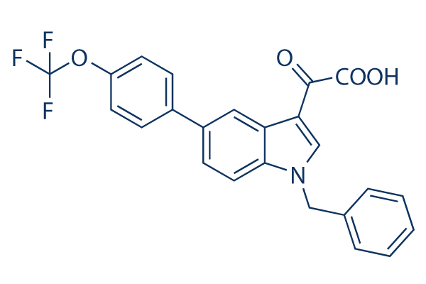
| Smiles | C1=CC=C(C=C1)CN2C=C(C3=C2C=CC(=C3)C4=CC=C(C=C4)OC(F)(F)F)C(=O)C(=O)O | ||
| 保管 | |||
|
In vitro |
DMSO : 88 mg/mL ( (200.28 mM); 吸湿したDMSOは溶解度を減少させます。新しいDMSOをご使用ください。) Ethanol : 7 mg/mL Water : Insoluble |
モル濃度計算器 |
|
in vivo Add solvents to the product individually and in order. |
投与溶液組成計算機 | |||||
実験計算
投与溶液組成計算機(クリア溶液)
ステップ1:実験データを入力してください。(実験操作によるロスを考慮し、動物数を1匹分多くして計算・調製することを推奨します)
mg/kg
g
μL
匹
ステップ2:投与溶媒の組成を入力してください。(ロット毎に適した溶解組成が異なる場合があります。詳細については弊社までお問い合わせください)
% DMSO
%
% Tween 80
% ddH2O
%DMSO
%
計算結果:
投与溶媒濃度: mg/ml;
DMSOストック溶液調製方法: mg 試薬を μL DMSOに溶解する(濃度 mg/mL, 注:濃度が当該ロットのDMSO溶解度を超える場合はご連絡ください。 )
投与溶媒調製方法:Take μL DMSOストック溶液に μL PEG300,を加え、完全溶解後μL Tween 80,を加えて完全溶解させた後 μL ddH2O,を加え完全に溶解させます。
投与溶媒調製方法:μL DMSOストック溶液に μL Corn oil,を加え、完全溶解。
注意:1.ストック溶液に沈殿、混濁などがないことをご確認ください;
2.順番通りに溶剤を加えてください。次のステップに進む前に溶液に沈殿、混濁などがないことを確認してから加えてください。ボルテックス、ソニケーション、水浴加熱など物理的な方法で溶解を早めることは可能です。
技術サポート
ストックの作り方、阻害剤の保管方法、細胞実験や動物実験の際に注意すべき点など、製品を取扱う時に問い合わせが多かった質問に対しては取扱説明書でお答えしています。
他に質問がある場合は、お気軽にお問い合わせください。
* 必須
納期 国内在庫品:受注日の翌日(15時までの受注分) *北海道、九州、沖縄への配送は受注日より2日以上 を要する場合あり 海外在庫品:受注後1〜2週間