
- 阻害剤
- 研究分野別
- PI3K/Akt/mTOR
- Epigenetics
- Methylation
- Immunology & Inflammation
- Protein Tyrosine Kinase
- Angiogenesis
- Apoptosis
- Autophagy
- ER stress & UPR
- JAK/STAT
- MAPK
- Cytoskeletal Signaling
- Cell Cycle
- TGF-beta/Smad
- 化合物ライブラリー
- 抗体
- 新製品
- お問い合わせ
GDC-0152
GDC-0152は、XIAP-BIR3、ML-IAP-BIR3、cIAP1-BIR3、およびcIAP2-BIR3の強力なアンタゴニストであり、無細胞アッセイにおいてKiがそれぞれ28 nM、14 nM、17 nM、43 nMでした。cIAP1-BIR2およびcIAP2-BIR2に対してはより低い親和性を示します。フェーズ1。
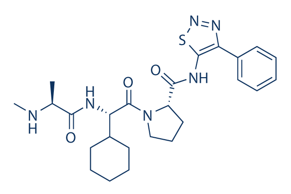
CAS No. 873652-48-3
文献中Selleckの製品使用例(22)
カスタマーフィードバック3例
製品安全説明書
現在のバッチを見る:
純度:
99.8%
99.8
GDC-0152関連製品
シグナル伝達経路
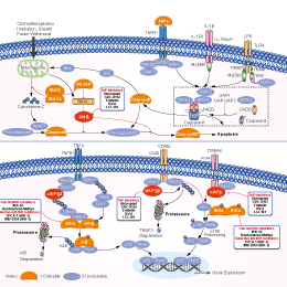
IAP阻害剤の選択性比較
Cell Data
| Cell Lines | Assay Type | Concentration | Incubation Time | 活性情報 | PMID |
|---|---|---|---|---|---|
| MDA-MB-231 cells | Cytotoxicity assay | 72 h | Cytotoxicity against human MDA-MB-231 cells assessed as decrease in cell viability after 72 hrs by CellTiter-Glo luminescent assay | ||
| A2058 cells | Function assay | 15 mins | Induction of proteasomal degradation of cIAP1 in human A2058 cells after 15 mins by immunoblotting | ||
| SK-MEL28 cells | Function assay | 0.5 μM | Inhibition of ML-IAP binding to Smax expressed in and zVAd treated human SK-MEL28 cells at 0.5 uM by immunoprecipitation | ||
| HEK293T cells | Function assay | 1-50 μM | 2 h | Inhibition of Flag-tagged XIAP BIR3 domain binding to cIAP1 expressed in human HEK293T cells at 1 to 50 uM after 2 hrs by immunoprecipitation | |
| 他の多くの細胞株試験データをご覧になる場合はこちらをクリックして下さい | |||||
生物活性
| 製品説明 | GDC-0152は、XIAP-BIR3、ML-IAP-BIR3、cIAP1-BIR3、およびcIAP2-BIR3の強力なアンタゴニストであり、無細胞アッセイにおいてKiがそれぞれ28 nM、14 nM、17 nM、43 nMでした。cIAP1-BIR2およびcIAP2-BIR2に対してはより低い親和性を示します。フェーズ1。 | ||||||||||
|---|---|---|---|---|---|---|---|---|---|---|---|
| Targets |
|
| In Vitro | ||||
| In vitro | GDC-0152 can block protein−protein interactions that involve IAP proteins and pro-apoptotic molecules. Using transiently transfected HEK293T cells, this compound is shown to disrupt XIAP binding to partially processed caspase-9 and to disrupt the association of ML-IAP, cIAP1, and cIAP2 with Smac. In melanoma SK-MEL28 cells, the endogenous association of ML-IAP and Smac is effectively also abolished by this chemical. It leads to a decrease in cell viability in the MDA-MB-231 breast cancer cell line, while having no effect on normal human mammary epithelial cells (HMEC). This compound is found to activate caspases 3 and 7 in a dose- and time-dependent manner. It is shown to induce rapid degradation of cIAP1 in A2058 melanoma cells. It effectively induces degradation of cIAP1 at concentrations as low as 10 nM, consistent with its affinity for cIAP1. | |||
|---|---|---|---|---|
| Kinase Assay | Fluorescence polarization-based competition assay | |||
| Inhibition constants ( Ki ) for the antagonists are determined by addition of the IAP protein constructs to wells containing serial dilutions of the antagonists or the peptide AVPW, and the Hid-FAM probe or AVP-diPhe-FAM probe, as appropriate, in the polarization buffer. Samples are read after a 30-minute incubation. Fluorescence polarization values are plotted as a function of the antagonist concentration, and the IC50 values are obtained by fitting the data to a 4-parameter equation using software. Ki values for the antagonists are determined from the IC50 valued. | ||||
| 細胞実験 | 細胞株 | MDA-MB-231, Normal human mammary epithelial cells (HMECs) | ||
| 濃度 | ~1 μM | |||
| 反応時間 | 72 h | |||
| 実験の流れ | MDA-MB-231 breast carcinoma cells and HMECs are treated with the indicated concentrations of GDC-0152. Cell death is assessed using the CellTiter-Glo luminescent cell viability assay 72 h following the start of treatment with this compound. | |||
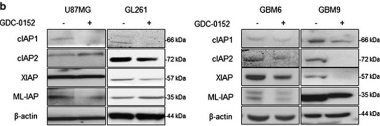
| 実験結果図 | Methods | Biomarkers | 結果図 | PMID |
| Western blot | cIAP1 / cIAP2 / XIAP / ML-IAP |
|
27490930 | |
| In Vivo | ||
| In Vivo | GDC-0152 has moderate predicted hepatic clearance based on metabolic stability assays conducted using human liver microsomes. Plasma−protein binding of this compound is moderate and comparable among mice (88−91%), rats (89−91%), dogs (81−90%), monkeys (76−85%), and humans (75−83%) over the range of concentrations investigated (0.1−100 μM); higher plasma−protein binding is observed in rabbits (95−96%). It does not preferentially distribute to red blood cells with blood−plasma partition ratios ranging from 0.6 to 1.1 in all species tested. The pharmacokinetics for this chemical is achieved with a C max of 53.7 μM and AUC of 203.5 h•μM. | |
|---|---|---|
| 動物実験 | 動物モデル | human-tumor xenograft mouse models of MDA-MB-231 breast cancer |
| 投与量 | 10, 50, or 100 mg/kg | |
| 投与経路 | oral gavage | |
| NCT Number | Recruitment | Conditions | Sponsor/Collaborators | Start Date | Phases |
|---|---|---|---|---|---|
| NCT00977067 | Terminated | Solid Cancers |
Genentech Inc. |
June 2007 | Phase 1 |
|
化学情報
| 分子量 | 498.64 | 化学式 | C25H34N6O3S |
| CAS No. | 873652-48-3 | SDF | Download GDC-0152 SDFをダウンロードする |
| Smiles | CC(C(=O)NC(C1CCCCC1)C(=O)N2CCCC2C(=O)NC3=C(N=NS3)C4=CC=CC=C4)NC | ||
| 保管 | |||
|
In vitro |
DMSO : 99 mg/mL ( (198.54 mM); 吸湿したDMSOは溶解度を減少させます。新しいDMSOをご使用ください。) Ethanol : 99 mg/mL Water : Insoluble |
モル濃度計算器 |
|
in vivo Add solvents to the product individually and in order. |
投与溶液組成計算機 | |||||
実験計算
投与溶液組成計算機(クリア溶液)
ステップ1:実験データを入力してください。(実験操作によるロスを考慮し、動物数を1匹分多くして計算・調製することを推奨します)
mg/kg
g
μL
匹
ステップ2:投与溶媒の組成を入力してください。(ロット毎に適した溶解組成が異なる場合があります。詳細については弊社までお問い合わせください)
% DMSO
%
% Tween 80
% ddH2O
%DMSO
%
計算結果:
投与溶媒濃度: mg/ml;
DMSOストック溶液調製方法: mg 試薬を μL DMSOに溶解する(濃度 mg/mL, 注:濃度が当該ロットのDMSO溶解度を超える場合はご連絡ください。 )
投与溶媒調製方法:Take μL DMSOストック溶液に μL PEG300,を加え、完全溶解後μL Tween 80,を加えて完全溶解させた後 μL ddH2O,を加え完全に溶解させます。
投与溶媒調製方法:μL DMSOストック溶液に μL Corn oil,を加え、完全溶解。
注意:1.ストック溶液に沈殿、混濁などがないことをご確認ください;
2.順番通りに溶剤を加えてください。次のステップに進む前に溶液に沈殿、混濁などがないことを確認してから加えてください。ボルテックス、ソニケーション、水浴加熱など物理的な方法で溶解を早めることは可能です。
技術サポート
ストックの作り方、阻害剤の保管方法、細胞実験や動物実験の際に注意すべき点など、製品を取扱う時に問い合わせが多かった質問に対しては取扱説明書でお答えしています。
他に質問がある場合は、お気軽にお問い合わせください。
* 必須
納期 国内在庫品:受注日の翌日(15時までの受注分) *北海道、九州、沖縄への配送は受注日より2日以上 を要する場合あり 海外在庫品:受注後1〜2週間