
- 阻害剤
- 研究分野別
- PI3K/Akt/mTOR
- Epigenetics
- Methylation
- Immunology & Inflammation
- Protein Tyrosine Kinase
- Angiogenesis
- Apoptosis
- Autophagy
- ER stress & UPR
- JAK/STAT
- MAPK
- Cytoskeletal Signaling
- Cell Cycle
- TGF-beta/Smad
- 化合物ライブラリー
- 抗体
- 新製品
- お問い合わせ
LCL161
LCL-161は、低分子のSMAC (second mitochondrial activator of caspase)ミメティックであり、複数のIAP(XIAP、c-IAPなど)に強力に結合し、それらを阻害します。
CAS No. 1005342-46-0
文献中Selleckの製品使用例(49)
製品安全説明書
現在のバッチを見る:
純度:
99.99%
99.99
LCL161関連製品
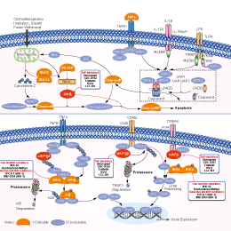
シグナル伝達経路
IAP阻害剤の選択性比較
Cell Data
| Cell Lines | Assay Type | Concentration | Incubation Time | 活性情報 | PMID |
|---|---|---|---|---|---|
| LOX | Function assay | 100 mg/kg | 8 hrs | Plasma concentration in nude mouse xenografted with human LOX cells at 100 mg/kg, po measured at 8 hrs, Cp = 3.3 μM. | 24093940 |
| H460 | Function assay | 3.3 uM | 5 days | Potentiation of conatumumab-induced cytotoxicity against human H460 cells at 3.3 uM after 5 days by MTS assay | 24083782 |
| LS180 | Function assay | 3.3 uM | 5 days | Potentiation of conatumumab-induced cytotoxicity against human LS180 cells at 3.3 uM after 5 days by MTS assay | 24083782 |
| LOX | Function assay | 3.3 uM | 3 days | Potentiation of conatumumab-induced cytotoxicity against human LOX cells at 3.3 uM after 3 days by MTS assay | 24083782 |
| SW620 | Function assay | 3.3 uM | 5 days | Potentiation of conatumumab-induced cytotoxicity against human SW620 cells at 3.3 uM after 5 days by MTS assay | 24083782 |
| SW620 | Function assay | 1.1 uM | 5 days | Potentiation of conatumumab-induced cytotoxicity against human SW620 cells at 1.1 uM after 5 days by MTS assay | 24083782 |
| HCT15 | Function assay | 3.3 uM | 5 days | Potentiation of conatumumab-induced cytotoxicity against human HCT15 cells at 3.3 uM after 5 days by MTS assay | 24083782 |
| BxPC3 | Function assay | 3.3 uM | 5 days | Potentiation of conatumumab-induced cytotoxicity against human BxPC3 cells at 3.3 uM after 5 days by MTS assay | 24083782 |
| MDA-MB-231 | Function assay | 2.5 to 10 uM | 19 hrs | Binding affinity to cIAP1 BIR3 domain in human MDA-MB-231 cells assessed as increase in TNFalpha level at 2.5 to 10 uM after 19 hrs by ELISA | 24083782 |
| LOX | Function assay | 100 mg/kg | 8 hrs | Potentiation of conatumumab-induced cIAP1 degradation in human LOX cells xenografted in mouse at 100 mg/kg, po after 8 hrs by Western blotting analysis | 24083782 |
| LOX | Function assay | 100 mg/kg | 8 hrs | Drug uptake in tumor of nude mouse xenografted with human LOX cells at 100 mg/kg, po measured at 8 hrs, Drug uptake = 18.4 μM. | 24093940 |
| SW620 | Function assay | 2.5 uM | 5 days | Induction of sensitization of human SW620 cells to conatumumab-induced apoptosis assessed as cell viability at 2.5 uM after 5 days by MTS assay | 24093940 |
| LOX | Function assay | 100 mg/kg | 8 hrs | In vivo inhibition of XIAP BIR2 domain in human LOX cells xenografted in nude mouse assessed as potentiation of conatumumab-induced caspase 3/7 activity at 100 mg/kg, po after 8 hrs by Western blot analysis | 24093940 |
| LOX | Function assay | 100 mg/kg | 8 hrs | In vivo inhibition of XIAP BIR2 domain in human LOX cells xenografted in nude mouse assessed as increase in caspase 3/7 activity at 100 mg/kg, po after 8 hrs by Western blot analysis | 24093940 |
| MDA-MB-231 | Function assay | 0.37 to 3.3 uM | 19 hrs | Inhibition of cIAP1/2 in human MDA-MB-231 cells assessed as induction of TNFalpha level at 0.37 to 3.3 uM after 19 hrs by ELISA | 24093940 |
| CHL1 | Function assay | 0.4 to 10 uM | 28 hrs | Inhibition of cIAP1 in human CHL1 cells at 0.4 to 10 uM after 28 hrs by Western blot analysis | 24093940 |
| SKMES1 | Function assay | 2.5 uM | 5 days | Potentiation of conatumumab-induced apoptosis in human SKMES1 cells at 2.5 uM after 5 days by MTS assay | 24093940 |
| Capan1 | Function assay | 2.5 uM | 5 days | Potentiation of conatumumab-induced apoptosis in human Capan1 cells at 2.5 uM after 5 days by MTS assay | 24093940 |
| AGS | Function assay | 2.5 uM | 5 days | Induction of sensitization of human AGS cells to conatumumab-induced apoptosis assessed as cell viability at 2.5 uM after 5 days by MTS assay | 24093940 |
| U118MG | Function assay | 2.5 uM | 5 days | Potentiation of conatumumab-induced apoptosis in human U118MG cells at 2.5 uM after 5 days by MTS assay | 24093940 |
| PC3 | Function assay | 2.5 uM | 5 days | Induction of sensitization of human PC3 cells to conatumumab-induced apoptosis assessed as cell viability at 2.5 uM after 5 days by MTS assay | 24093940 |
| MDA-MB-231 | Antitumor assay | 30 mg/kg | 24 days | Antitumor activity against human MDA-MB-231 cells xenografted in Balb/c SCID mouse assessed as tumor growth inhibition at 30 mg/kg administered via oral gavage for 24 days | 28492317 |
| A549 | Function assay | 1 uM | 3 hrs | Induction of cIAP2 degradation in human A549 cells assessed as reduction in cIAP2 protein level at 1 uM incubated for 3 hrs by Western blot analysis | 31550155 |
| SK-MEL-28 | Function assay | 1 uM | 3 hrs | Induction of cIAP1 degradation in human SK-MEL-28 cells assessed as reduction in cIAP1 protein level at 1 uM incubated for 3 hrs by Western blot analysis | 31550155 |
| SK-MEL-28 | Function assay | 1 uM | 3 hrs | Induction of cIAP2 degradation in human SK-MEL-28 cells assessed as reduction in cIAP2 protein level at 1 uM incubated for 3 hrs by Western blot analysis | 31550155 |
| HEK293T | Function assay | 10 uM | 6 hrs | Covalent binding affinity to HA-BIR3 domain of XIAP (unknown origin) expressed in HEK293T cells assessed as increase in band intensity at 10 uM incubated for 6 hrs by Western blot analysis | 31550155 |
| A549 | Function assay | 1 uM | 3 hrs | Induction of cIAP1 degradation in human A549 cells assessed as reduction in cIAP1 protein level at 1 uM incubated for 3 hrs by Western blot analysis | 31550155 |
| LOX | Function assay | 8 hrs | Potentiation of conatumumab-induced caspase 3/7 activation in human LOX cells xenografted in mouse after 8 hrs by fluorescence assay | 24083782 | |
| SW620 | Function assay | 5 days | Induction of sensitization of human SW620 cells to conatumumab-induced apoptosis assessed as cell viability after 5 days by MTS assay, EC90 = 6.66 μM. | 24093940 | |
| MDA-MB-231 | Function assay | 2 hrs | Induction of cIAP1 degradation in human MDA-MB-231 cells after 2 hrs, EC50 = 0.0004 μM. | 28492317 | |
| MDA-MB-231 | Cytotoxicity assay | 72 hrs | Cytotoxicity against human MDA-MB-231 cells assessed as decrease in cell proliferation after 72 hrs by alamar blue assay, EC50 = 0.0078 μM. | 28492317 | |
| HEK293 | Function assay | 2 hrs | Inhibition of full length FLAG-tagged XIAP (unknown origin) interaction with full length untagged caspase-9 expressed in HEK293 cells after 2 hrs by immunoprecipitation assay, EC50 = 0.035 μM. | 28492317 | |
| MDA-MB-231 | Function assay | 2 hrs | Induction of intracellular cIAP1 degradation in human MDA-MB-231 cells after 2 hrs, IC50 = 0.0004 μM. | 30091600 | |
| MDA-MB-231 | Antiproliferative assay | 72 hrs | Antiproliferative activity against human MDA-MB-231 cells after 72 hrs by Alamar blue assay, IC50 = 0.0078 μM. | 30091600 | |
| HEK293 | Function assay | 2 hrs | Inhibition of full length FLAG-tagged XIAP (unknown origin) interaction with full length untagged caspase-9 expressed in HEK293 cells after 2 hrs by immunoprecipitation assay, IC50 = 0.035 μM. | 30091600 | |
| SKOV3 | Apoptosis assay | 48 hrs | Induction of apoptosis in human SKOV3 cells assessed caspase-3 activation after 48 hrs by IncuCyte S3 live-cell analysis, EC50 = 0.001 μM. | 31095386 | |
| SKOV3 | Apoptosis assay | 24 hrs | Induction of apoptosis in human SKOV3 cells assessed caspase-3 activation after 24 hrs by IncuCyte S3 live-cell analysis, EC50 = 0.003 μM. | 31095386 | |
| BL21(DE3) | Function assay | 2 hrs | Displacement of biotinylated AVPF from N-terminal His tagged recombinant human XIAP-BIR3 domain (253 to 347 residues) expressed in Escherichia coli BL21(DE3) cells incubated for 2 hrs by DELFIA, IC50 = 0.048 μM. | 31095386 | |
| BL21(DE3) | Function assay | 8 hrs | Displacement of biotinylated AVPF from N-terminal His tagged recombinant human XIAP-BIR3 domain (253 to 347 residues) expressed in Escherichia coli BL21(DE3) cells incubated for 8 hrs by DELFIA, IC50 = 0.053 μM. | 31095386 | |
| CCRF-CEM | Antiproliferative assay | Antiproliferative activity against human CCRF-CEM cells, GI50 = 0.25 μM. | 28435526 | ||
| 他の多くの細胞株試験データをご覧になる場合はこちらをクリックして下さい | |||||
生物活性
| 製品説明 | LCL-161は、低分子のSMAC (second mitochondrial activator of caspase)ミメティックであり、複数のIAP(XIAP、c-IAPなど)に強力に結合し、それらを阻害します。 | ||
|---|---|---|---|
| Targets |
|
| In Vitro | ||||
| In vitro |
LCL161 binds to inhibitors of apoptosis proteins (IAPs) with high affinity and initiates the destruction of cIAP1 and cIAP2, which further induces apoptosis via caspase activation. This compound modestly inhibits the growth of FLT3-ITD-expressing cells when administered alone, with an IC50 ranging from ~0.5 μM (Ba/F3-FLT3-ITD cells) to ~4 μM (MOLM13-luc+ cells). The potency of this agent against the D835Y mutant is observed to be considerably higher, with an IC50 of ~50 nM when tested against Ba/F3-D835Y cells. Treatment of MOLM13-luc+ cells with a combination of this compound and PKC412 leads to significantly more killing of cells than either agent alone, with Calcusyn combination indices suggestive of synergy. PKC412 and this chemical induces apoptosis of MOLM13-luc+ cells. The combination of PKC412 and this agent leads to a higher induction of apoptosis than either agent alone. It is able to override stromal-mediated rescue of mutant FLT3-expressing cells through positive combination with PKC412. This compound inhibits the growth of Ba/F3.p210 cells with an IC50 of ~100 nM. It also has demonstrated activity against drug-resistant cells expressing point mutations in the target proteins. This agent at 1000 nM is able to mostly or completely kill Ba/F3-derived cell lines conferring resistance to PKC412, which express FLT3-ITD harboring point mutations in the ATP-binding pocket of FLT3. It also shows activity at concentrations ranging from 100 to 1000 nM against Ba/F3 cells expressing various. This compound is evaluated against the 23 cell lines in the Pediatric Preclinical Testing Program (PPTP) in vitro panel using 96 hr. It achieves 50% growth inhibition against only 3 of the 23 tested PPTP cell lines under concentration of 10 μM. The three cell lines includes two T-cell ALL cell lines (COG-LL-317 and CCRF-CEM) and an anaplastic large cell lymphoma cell line (Karpas-299), with CCRF-CEM and Karpas-299 showing the lowest relative IC50 values (0.25 and 1.6 μM, respectively). It shows immunomodulatory properties on human immune subsets. T lymphocytes treated with this compound demonstrates significantly enhanced cytokine secretion upon activation, with little effect on CD4 and CD8 T-cell survival or proliferation. This agent treatment of peripheral blood mononuclear cells significantly enhances priming of naïve T cells with synthetic peptides in vitro. Myeloid dendritic cells undergoes phenotypic maturation upon this chemical and demonstrates a reduced capacity to cross-present a tumor antigen-based vaccine. These effects are potentially mediated through an observed activation of the canonical and non-canonical NF-κB pathways, following this compound with a resulting upregulation of anti-apoptotic molecules. |
|||
|---|---|---|---|---|
| 細胞実験 | 細胞株 | Human T-cell ALL cell lines COG-LL-317 | ||
| 濃度 | ~10 μM | |||
| 反応時間 | 96 hours | |||
| 実験の流れ | In vitro testing is performed using DIMSCAN |
|||
| 実験結果図 | Methods | Biomarkers | 結果図 | PMID |
| Western blot | cIAP1 / cIAP2 / XIAP / surivivin |
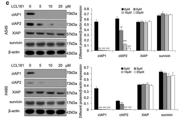
|
27737687 | |
| Growth inhibition assay | Cell viability |
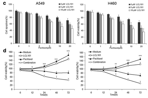
|
27737687 | |
| In Vivo | ||
| In Vivo |
LCL161 significantly enhances the ability of PKC412 to inhibit the growth of Ba/F3-FLT3-ITD-luc+ cells in vivo. This compound is also shown to positively combine with the standard chemotherapeutic agents, Ara-c and doxorubicin, against FLT3-ITD-expressing cells and against D835Y-expressing cells. There is an additive effect achieved by combining both this chemical in suppressing leukemia growth. This agent (100 mg/kg) enhances in vivo effects of high-moderate doses on leukemia burden in mice. This compound is tested against the Pediatric Preclinical Testing Program (PPTP) in vivo panels (30 or 75 mg/kg [solid tumors] or 100 mg/kg [ALL]) administered orally twice in a week. It induces significant differences in EFS distribution in approximately one-third of solid tumor xenografts (osteosarcoma and glioblastoma), but not in ALL xenografts. No objective tumor responses are observed. In vivo this chemical demonstrates limited single agent activity against the pediatric preclinical models studied. |
|
|---|---|---|
| 動物実験 | 動物モデル | CB17SC scid−/− female mice |
| 投与量 | 30 mg/kg | |
| 投与経路 | o.g. | |
| NCT Number | Recruitment | Conditions | Sponsor/Collaborators | Start Date | Phases |
|---|---|---|---|---|---|
| NCT03111992 | Completed | Multiple Myeloma |
Novartis Pharmaceuticals|Novartis |
December 18 2017 | Phase 1 |
| NCT01934634 | Unknown status | Metastatic Pancreatic Cancer |
US Oncology Research|Novartis Pharmaceuticals|Delta Clinical Research LLC |
March 2014 | Phase 1 |
| NCT01968915 | Completed | Neoplasms |
Novartis Pharmaceuticals|Novartis |
November 2013 | Phase 1 |
| NCT01617668 | Completed | Breast Cancer |
Novartis Pharmaceuticals|Novartis |
August 2012 | Phase 2 |
|
化学情報
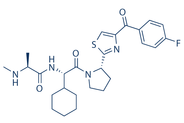
| 分子量 | 500.63 | 化学式 | C26H33FN4O3S |
| CAS No. | 1005342-46-0 | SDF | Download LCL161 SDFをダウンロードする |
| Smiles | CC(C(=O)NC(C1CCCCC1)C(=O)N2CCCC2C3=NC(=CS3)C(=O)C4=CC=C(C=C4)F)NC | ||
| 保管 | |||
|
In vitro |
DMSO : 100 mg/mL ( (199.74 mM); 吸湿したDMSOは溶解度を減少させます。新しいDMSOをご使用ください。) Ethanol : 100 mg/mL Water : Insoluble |
モル濃度計算器 |
|
in vivo Add solvents to the product individually and in order. |
投与溶液組成計算機 | |||||
実験計算
投与溶液組成計算機(クリア溶液)
ステップ1:実験データを入力してください。(実験操作によるロスを考慮し、動物数を1匹分多くして計算・調製することを推奨します)
mg/kg
g
μL
匹
ステップ2:投与溶媒の組成を入力してください。(ロット毎に適した溶解組成が異なる場合があります。詳細については弊社までお問い合わせください)
% DMSO
%
% Tween 80
% ddH2O
%DMSO
%
計算結果:
投与溶媒濃度: mg/ml;
DMSOストック溶液調製方法: mg 試薬を μL DMSOに溶解する(濃度 mg/mL, 注:濃度が当該ロットのDMSO溶解度を超える場合はご連絡ください。 )
投与溶媒調製方法:Take μL DMSOストック溶液に μL PEG300,を加え、完全溶解後μL Tween 80,を加えて完全溶解させた後 μL ddH2O,を加え完全に溶解させます。
投与溶媒調製方法:μL DMSOストック溶液に μL Corn oil,を加え、完全溶解。
注意:1.ストック溶液に沈殿、混濁などがないことをご確認ください;
2.順番通りに溶剤を加えてください。次のステップに進む前に溶液に沈殿、混濁などがないことを確認してから加えてください。ボルテックス、ソニケーション、水浴加熱など物理的な方法で溶解を早めることは可能です。
技術サポート
ストックの作り方、阻害剤の保管方法、細胞実験や動物実験の際に注意すべき点など、製品を取扱う時に問い合わせが多かった質問に対しては取扱説明書でお答えしています。
他に質問がある場合は、お気軽にお問い合わせください。
* 必須
納期 国内在庫品:受注日の翌日(15時までの受注分) *北海道、九州、沖縄への配送は受注日より2日以上 を要する場合あり 海外在庫品:受注後1〜2週間