
- 阻害剤
- 研究分野別
- PI3K/Akt/mTOR
- Epigenetics
- Methylation
- Immunology & Inflammation
- Protein Tyrosine Kinase
- Angiogenesis
- Apoptosis
- Autophagy
- ER stress & UPR
- JAK/STAT
- MAPK
- Cytoskeletal Signaling
- Cell Cycle
- TGF-beta/Smad
- 化合物ライブラリー
- 抗体
- 新製品
- お問い合わせ
Vandetanib (ZD6474)
別名:ZD6474
バンデタニブは、細胞フリーアッセイにおいてIC50が40 nMの強力なVEGFR2阻害剤です。また、VEGFR3とEGFRもそれぞれIC50が110 nMおよび500 nMで阻害します。PDGFRβ、Flt1、Tie-2、FGFR1には感受性がなく、IC50は1.1-3.6 μMです。MEK、CDK2、c-Kit、erbB2、FAK、PDK1、Akt、IGF-1Rに対しては10 μMを超えるIC50で活性がありません。バンデタニブ(ZD6474)は、reactive oxygen species (ROS)のレベルを上昇させることにより、apoptosisを増加させ、autophagyを誘導します。
CAS No. 443913-73-3
文献中Selleckの製品使用例(113)
製品安全説明書
現在のバッチを見る:
純度:
99.99%
99.99
Vandetanib (ZD6474)関連製品
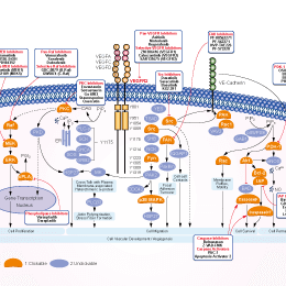
シグナル伝達経路
VEGFR阻害剤の選択性比較
Cell Data
| Cell Lines | Assay Type | Concentration | Incubation Time | 活性情報 | PMID |
|---|---|---|---|---|---|
| PCI-15B | Function Assay | 1 μM | 24 h | downregulates VEGF production | 22307735 |
| UM-22A | Function Assay | 1 μM | 24 h | downregulates VEGF production | 22307735 |
| PCI-37A | Function Assay | 1 μM | 24 h | downregulates VEGF production | 22307735 |
| PCI-15B | Function Assay | 0-10 μM | 24 h | inhibits the activation of the EGFR tyrosine kinase and also decreases the expression of phosphorylated forms of the downstream signaling elements, STAT3 and MAPK | 22307735 |
| UM-22B | Function Assay | 0-10 μM | 24 h | inhibits the activation of the EGFR tyrosine kinase and also decreases the expression of phosphorylated forms of the downstream signaling elements, STAT3 and MAPK | 22307735 |
| UM-22A | Function Assay | 0-10 μM | 24 h | inhibits the activation of the EGFR tyrosine kinase and also decreases the expression of phosphorylated forms of the downstream signaling elements, STAT3 and MAPK | 22307735 |
| SCC-25 | Growth Inhibition Assay | 0-6 μM | 72 h | inhibits cell growth in a dose dependent manner | 22307735 |
| PCI-15B | Growth Inhibition Assay | 0-6 μM | 72 h | inhibits cell growth in a dose dependent manner | 22307735 |
| PCI-37B | Growth Inhibition Assay | 0-6 μM | 72 h | inhibits cell growth in a dose dependent manner | 22307735 |
| PCI-37A | Growth Inhibition Assay | 0-6 μM | 72 h | inhibits cell growth in a dose dependent manner | 22307735 |
| UM-22B | Growth Inhibition Assay | 0-6 μM | 72 h | inhibits cell growth in a dose dependent manner | 22307735 |
| UM-22A | Growth Inhibition Assay | 0-6 μM | 72 h | inhibits cell growth in a dose dependent manner | 22307735 |
| HAK1-B | Function Assay | 1/5/10 μM | 1 h | suppresses EGFR phosphorylation | 22611027 |
| HUVECs | Function Assay | 1/5/10 μM | 1 h | significantly inhibits VEGFR-2 phosphorylation | 22611027 |
| U87MG | Function Assay | 4 μℳ | 2/6/12 h | suppresses basal levels of phosphorylation of S6 (S235/236), 4E-BP1 (T37/46), and Akt (S473) in a time-dependent manner | 23799852 |
| U251 | Function Assay | 4 μℳ | 2/6/12 h | suppresses basal levels of phosphorylation of S6 (S235/236), 4E-BP1 (T37/46), and Akt (S473) in a time-dependent manner | 23799852 |
| U87MG | Function Assay | 2/4/8 μℳ | 6/12/24 h | increases the LC3-II level in a time-dependent and dose-dependent manner | 23799852 |
| U251 | Function Assay | 2/4/8 μℳ | 6/12/24 h | increases the LC3-II level in a time-dependent and dose-dependent manner | 23799852 |
| MDA-MB-468 | Growth Inhibition Assay | 1 nM-100 μM | 48 h | inhibits cell growth in a dose dependent manner | 24138843 |
| T-47-D | Growth Inhibition Assay | 1 nM-100 μM | 48 h | inhibits cell growth in a dose dependent manner | 24138843 |
| MDA-MB-231 | Growth Inhibition Assay | 1 nM-100 μM | 48 h | inhibits cell growth in a dose dependent manner | 24138843 |
| ZR-75-1 | Growth Inhibition Assay | 1 nM-100 μM | 48 h | inhibits cell growth in a dose dependent manner | 24138843 |
| MCF-7 | Growth Inhibition Assay | 1 nM-100 μM | 48 h | inhibits cell growth in a dose dependent manner | 24138843 |
| HMEpC | Growth Inhibition Assay | 1 nM-100 μM | 48 h | inhibits cell growth in a dose dependent manner | 24138843 |
| SH-SY5Y | Function Assay | 5 μM | 48/72 h | suppresses expression of the CXCR4 and MMP14 protein | 24399074 |
| SK-N-SH | Function Assay | 5 μM | 48/72 h | suppresses expression of the CXCR4 and MMP14 protein | 24399074 |
| SH-SY5Y | Function Assay | 5 μM | 24/48/72 h | suppresses the expression of CXCR4 and MMP14 mRNA | 24399074 |
| SK-N-SH | Function Assay | 5 μM | 24/48/72 h | suppresses the expression of CXCR4 and MMP14 mRNA | 24399074 |
| SH-SY5Y | Function Assay | 5/10 μM | 48 h | inhibits human NB cell invasion | 24399074 |
| SK-N-SH | Function Assay | 5/10 μM | 48 h | inhibits human NB cell invasion | 24399074 |
| SH-SY5Y | Function Assay | 5/10 μM | 48 h | inhibits human NB cell migration | 24399074 |
| SK-N-SH | Function Assay | 5/10 μM | 48 h | inhibits human NB cell migration | 24399074 |
| SH-SY5Y | Function Assay | 1/5/10 μM | 48 h | inhibits RET phosphorylation | 24399074 |
| SK-N-SH | Function Assay | 1/5/10 μM | 48 h | inhibits RET phosphorylation | 24399074 |
| SH-SY5Y | Function Assay | 5/10/20 μM | 48 h | induces G1 phase cell cycle arrest | 24399074 |
| SK-N-SH | Function Assay | 5/10/20 μM | 48 h | induces G1 phase cell cycle arrest | 24399074 |
| SH-SY5Y | Apoptosisi Assay | 5/10/20 μM | 48 h | induces apoptosis dose dependently | 24399074 |
| SK-N-SH | Apoptosisi Assay | 5/10/20 μM | 48 h | induces apoptosis dose dependently | 24399074 |
| SH-SY5Y | Growth Inhibition Assay | 0.625-20 μM | 48 h | inhibits cell growth in a dose dependent manner | 24399074 |
| SK-N-SH | Growth Inhibition Assay | 0.625-20 μM | 48 h | inhibits cell growth in a dose dependent manner | 24399074 |
| SN179 | Function Assay | 500 nM | 16 h | increases basal migration | 25676691 |
| SN179 | Function Assay | 500 nM | 16 h | enhances the CXCL12 directed migration | 25676691 |
| SN186 | Function Assay | 500 nM | 16 h | increases CXCR4 expression significantly | 25676691 |
| SN179 | Function Assay | 500 nM | 16 h | increases CXCR4 expression significantly | 25676691 |
| 201T | Function Assay | 2.5 μM | 48 h | inhibits phospho-MAPK following EGF | 22258476 |
| 273T | Function Assay | 2.5 μM | 48 h | inhibits phospho-MAPK following EGF | 22258476 |
| A549 | Function Assay | 2.5 μM | 48 h | inhibits phospho-MAPK following EGF | 22258476 |
| 201T | Function Assay | 1/5/10 μM | 48 h | blocks the phosphorylation of Akt induced by VEGFC | 22258476 |
| HTB3 | Growth Inhibition Assay | 0-20 μM | 24 h | inhibits cell growth in a dose dependent manner | 19220256 |
| HT1376 | Growth Inhibition Assay | 0-20 μM | 24 h | inhibits cell growth in a dose dependent manner | 19220256 |
| RT4 | Growth Inhibition Assay | 0-20 μM | 24 h | inhibits cell growth in a dose dependent manner | 19220256 |
| J82 | Growth Inhibition Assay | 0-20 μM | 24 h | inhibits cell growth in a dose dependent manner | 19220256 |
| CRL1749 | Growth Inhibition Assay | 0-20 μM | 24 h | inhibits cell growth in a dose dependent manner | 19220256 |
| T24 | Growth Inhibition Assay | 0-20 μM | 24 h | inhibits cell growth in a dose dependent manner | 19220256 |
| SUP | Growth Inhibition Assay | 0-20 μM | 24 h | inhibits cell growth in a dose dependent manner | 19220256 |
| HTB9 | Growth Inhibition Assay | 0-20 μM | 24 h | inhibits cell growth in a dose dependent manner | 19220256 |
| ACC3 | Growth Inhibition Assay | 0-10 μM | 72 h | inhibits cell growth in a dose dependent manner | 18698025 |
| ACC2 | Growth Inhibition Assay | 0-10 μM | 72 h | inhibits cell growth in a dose dependent manner | 18698025 |
| ACCM | Growth Inhibition Assay | 0-10 μM | 72 h | inhibits cell growth in a dose dependent manner | 18698025 |
| ACC3 | Apoptosisi Assay | 0-10 μM | 72 h | induces apoptosis dose dependently | 18698025 |
| ACC2 | Apoptosisi Assay | 0-10 μM | 72 h | induces apoptosis dose dependently | 18698025 |
| ACCM | Apoptosisi Assay | 0-10 μM | 72 h | induces apoptosis dose dependently | 18698025 |
| CNE-1 | Growth Inhibition Assay | 0.1-25.6 μM | 48 h | IC50=3.6 μM | 17631646 |
| CNE-2 | Growth Inhibition Assay | 0.1-25.6 μM | 48 h | IC50=6.2 μM | 17631646 |
| C666-1 | Growth Inhibition Assay | 0.1-25.6 μM | 48 h | IC50=23.4 μM | 17631646 |
| CNE-1 | Growth Inhibition Assay | 0.1-25.6 μM | 72 h | IC50=2.3 μM | 17631646 |
| CNE-2 | Growth Inhibition Assay | 0.1-25.6 μM | 72 h | IC50=3.6 μM | 17631646 |
| C666-1 | Growth Inhibition Assay | 0.1-25.6 μM | 72 h | IC50=4.86 μM | 17631646 |
| CNE-1 | Function Assay | 6 μM | 24 h | delays G0/G1 cell cycle progression | 17631646 |
| CNE-2 | Function Assay | 6 μM | 24 h | delays G0/G1 cell cycle progression | 17631646 |
| C666-1 | Function Assay | 6 μM | 24 h | delays G0/G1 cell cycle progression | 17631646 |
| HT-29 | Antiproliferative assay | 10 uM | 72 hrs | Antiproliferative activity against human HT-29 cells at 10 uM after 72 hrs by MTS assay, IC50 = 4.2 μM. | 21353546 |
| EAhy926 | Antiproliferative assay | 10 uM | 72 hrs | Antiproliferative activity against human EAhy926 cells at 10 uM after 72 hrs by MTS assay, IC50 = 5.1 μM. | 21353546 |
| SCC-25 | Invasion Assay | 24 h | EC50=10 nM | 22307735 | |
| UM-22A | Invasion Assay | 24 h | EC50=0.3 nM | 22307735 | |
| PCI-37A | Invasion Assay | 24 h | EC50=1695 nM | 22307735 | |
| PCI-15B | Invasion Assay | 24 h | EC50=558 nM | 22307735 | |
| HuH-7 | Growth Inhibition Assay | 72 h | IC50 = 9.4 μmol/L | 22611027 | |
| KYN-2 | Growth Inhibition Assay | 72 h | IC50 = 8.1 μmol/L | 22611027 | |
| HUVECs | Growth Inhibition Assay | 72 h | IC50 = 7.1 μmol/L | 22611027 | |
| A-431 | Growth Inhibition Assay | 72 h | GI50=2.4 ± 0.3 μM | 24681205 | |
| NCTC-2544 | Growth Inhibition Assay | 72 h | GI50=4.6 ± 0.3 μM | 24681205 | |
| K-562 | Growth Inhibition Assay | 72 h | GI50=1.8 ± 0.1 μM | 24681205 | |
| Jurkat | Growth Inhibition Assay | 72 h | GI50=1.5 ± 0.2 μM | 24681205 | |
| UM-22B | Invasion Assay | 24 h | EC50=2424 nM | 22307735 | |
| PCI-37B | Invasion Assay | 24 h | EC50=1726 nM | 22307735 | |
| Hth83 | Growth Inhibition Assay | 72 h | IC50=3.30 ± 0.66 μM | 21220477 | |
| C643 | Growth Inhibition Assay | 72 h | IC50=3.65 ± 1.22 μM | 21220477 | |
| 8505C | Growth Inhibition Assay | 72 h | IC50=7.56 ± 1.13 μM | 21220477 | |
| Hth74 | Growth Inhibition Assay | 72 h | IC50=8.56 ± 1.01 μM | 21220477 | |
| SW1736 | Growth Inhibition Assay | 72 h | IC50=9.05 ± 0.55 μM | 21220477 | |
| Hth7 | Growth Inhibition Assay | 72 h | IC50=9.66 ± 0.38 μM | 21220477 | |
| Hth104 | Growth Inhibition Assay | 72 h | IC50=±16.98 ± NA μM | 21220477 | |
| EHMES-1 | Growth Inhibition Assay | 72 h | IC50=10.6 μM | 18364248 | |
| EHMES-10 | Growth Inhibition Assay | 72 h | IC50=0.3 μM | 18364248 | |
| 211H | Growth Inhibition Assay | 72 h | IC50=2.2 μM | 18364248 | |
| H28 | Growth Inhibition Assay | 72 h | IC50=1.8 μM | 18364248 | |
| H2052 | Growth Inhibition Assay | 72 h | IC50=8.0 μM | 18364248 | |
| H2452 | Growth Inhibition Assay | 72 h | IC50=5.5 μM | 18364248 | |
| TPC1 | Antiproliferative assay | 72 hrs | Antiproliferative activity against human TPC1 cells expressing RET/PCT1 after 72 hrs by [3H]thymidine incorporation assay, IC50 = 0.116 μM. | 20409618 | |
| Sf21 | Function assay | 15 mins | Inhibition of recombinant His-tagged human KDR expressed in insect Sf21 cells preincubated for 15 mins followed by substrate addition measured after 20 mins by HTRF assay, IC50 = 0.175 μM. | 26874741 | |
| BA/F3 | Function assay | 48 hrs | Inhibition of KIF5B/RET (unknown origin) expressed in mouse BA/F3 cells assessed as reduction in cell viability after 48 hrs by Cell titre glo-based luminescence assay, IC50 = 0.4 μM. | 26874741 | |
| BA/F3 | Function assay | 48 hrs | Inhibition of KDR (unknown origin) expressed in mouse BA/F3 cells assessed as reduction in cell viability after 48 hrs by Cell titre glo-based luminescence assay, IC50 = 0.63 μM. | 26874741 | |
| HL60 | Antiproliferative assay | 72 hrs | Antiproliferative activity against human HL60 cells after 72 hrs by MTT assay, IC50 = 1.492 μM. | 26995527 | |
| HT-29 | Antiproliferative assay | 72 hrs | Antiproliferative activity against human HT-29 cells after 72 hrs by MTT assay, IC50 = 1.925 μM. | 26995527 | |
| DU145 | Antiproliferative assay | 72 hrs | Antiproliferative activity against human DU145 cells after 72 hrs by MTT assay, IC50 = 1.974 μM. | 26995527 | |
| MGHU3 | Antiproliferative assay | 72 hrs | Antiproliferative activity against human MGHU3 cells after 72 hrs by CellTiter-Glo assay, IC50 = 2.5 μM. | 30309671 | |
| A549 | Antiproliferative assay | 72 hrs | Antiproliferative activity against human A549 cells after 72 hrs by CellTiter-Glo assay, IC50 = 2.5 μM. | 30309671 | |
| RT112 | Antiproliferative assay | 72 hrs | Antiproliferative activity against human RT112 cells after 72 hrs by CellTiter-Glo assay, IC50 = 2.5 μM. | 30309671 | |
| A549 | Antiproliferative assay | 72 hrs | Antiproliferative activity against human A549 cells after 72 hrs by MTT assay, IC50 = 2.63 μM. | 26995527 | |
| MCF7 | Antiproliferative assay | 72 hrs | Antiproliferative activity against human MCF7 cells after 72 hrs by MTT assay, IC50 = 3.536 μM. | 26995527 | |
| PANC1 | Antiproliferative assay | 72 hrs | Antiproliferative activity against human PANC1 cells after 72 hrs by MTT assay, IC50 = 4.107 μM. | 26995527 | |
| MCF7 | Antiproliferative assay | 48 hrs | Antiproliferative activity against human MCF7 cells measured after 48 hrs by MTT assay, IC50 = 11.83 μM. | 27688180 | |
| MCF7 | Cytotoxicity assay | 48 hrs | Cytotoxicity in human MCF7 cells assessed as inhibition of cell growth incubated for 48 hrs by MTT assay, IC50 = 16.52 μM. | 28942113 | |
| MCF7 | Antiproliferative assay | 48 hrs | Antiproliferative activity against human MCF7 cells after 48 hrs by MTT assay, IC50 = 18.5 μM. | 26741358 | |
| MCF7 | Antiproliferative assay | 48 hrs | Antiproliferative activity against human MCF7 cells after 48 hrs by MTT assay, IC50 = 18.5 μM. | 26475519 | |
| HT-29 | Antiproliferative assay | 48 hrs | Antiproliferative activity against human HT-29 cells measured after 48 hrs by MTT assay, IC50 = 18.95 μM. | 27688180 | |
| H460 | Antiproliferative assay | 48 hrs | Antiproliferative activity against human H460 cells measured after 48 hrs by MTT assay, IC50 = 37.1 μM. | 27688180 | |
| H1650 | Growth Inhibition Assay | IC50=3.5±1.2 μM | 23274758 | ||
| H2052 | Growth Inhibition Assay | IC50=1.07±0.04 μM | 21970874 | ||
| H2452 | Growth Inhibition Assay | IC50=3.52±1.13 μM | 21970874 | ||
| H28 | Growth Inhibition Assay | IC50=0.32±0.07 μM | 21970874 | ||
| MSTO-211H | Growth Inhibition Assay | IC50=1.42±0.03 μM | 21970874 | ||
| KDR15 | Function assay | Inhibitory activity against VEGF stimulated autophosphorylation of VEGFR2 expressed in KDR15 cells, IC50 = 0.015 μM. | 16302797 | ||
| Sf9 | Function assay | Inhibition of human recombinant histidine-tagged RET (700-1020) expressed in Sf9 cells by ELISA, IC50 = 0.097 μM. | 20409618 | ||
| HEK293 | Function assay | Inhibition of FGFR1/VEGFR2 chimeric construct expressed in HEK293 cells by ELISA, ED50 = 0.15 μM. | 19101155 | ||
| umbilical vein endothelial cells | Function assay | Inhibition of VEGF-induced proliferation of human umbilical vein endothelial cells, IC50 = 0.4 μM. | 15743202 | ||
| umbilical vein endothelial cells | Function assay | Inhibition of basic FGF-induced proliferation of human umbilical vein endothelial cells, IC50 = 1.2 μM. | 15743202 | ||
| 293 | Function assay | Inhibitory activity against VEGFR2 transiently transfected in 293 adenovirus transfected kidney cells by ELISA, IC50 = 1.66 μM. | 16275072 | ||
| 293 | Function assay | Inhibition of VEGFR2 in 293 adenovirus transfected kidney cells by cell-based ELISA assay, IC50 = 1.66 μM. | 16321531 | ||
| HEK293 | Function assay | Inhibition of VEGFR2 phosphorylation in HEK293 cells by cell-based ELISA, IC50 = 1.66 μM. | 16460936 | ||
| CHO | Function assay | Inhibition of VEGFR induced autophosphorylation of human Vascular endothelial growth factor receptor 2 (VEGFR2) transfected in CHO cells, IC50 = 2.673 μM. | 12477352 | ||
| A673 | qHTS assay | qHTS of pediatric cancer cell lines to identify multiple opportunities for drug repurposing: Primary screen for A673 cells | 29435139 | ||
| DAOY | qHTS assay | qHTS of pediatric cancer cell lines to identify multiple opportunities for drug repurposing: Primary screen for DAOY cells | 29435139 | ||
| BT-37 | qHTS assay | qHTS of pediatric cancer cell lines to identify multiple opportunities for drug repurposing: Primary screen for BT-37 cells | 29435139 | ||
| RD | qHTS assay | qHTS of pediatric cancer cell lines to identify multiple opportunities for drug repurposing: Primary screen for RD cells | 29435139 | ||
| SK-N-SH | qHTS assay | qHTS of pediatric cancer cell lines to identify multiple opportunities for drug repurposing: Primary screen for SK-N-SH cells | 29435139 | ||
| MG 63 (6-TG R) | qHTS assay | qHTS of pediatric cancer cell lines to identify multiple opportunities for drug repurposing: Primary screen for MG 63 (6-TG R) cells | 29435139 | ||
| NB1643 | qHTS assay | qHTS of pediatric cancer cell lines to identify multiple opportunities for drug repurposing: Primary screen for NB1643 cells | 29435139 | ||
| OHS-50 | qHTS assay | qHTS of pediatric cancer cell lines to identify multiple opportunities for drug repurposing: Primary screen for OHS-50 cells | 29435139 | ||
| SK-N-SH | qHTS assay | qHTS of pediatric cancer cell lines to identify multiple opportunities for drug repurposing: Confirmatory screen for SK-N-SH cells | 29435139 | ||
| Rh41 | qHTS assay | qHTS of pediatric cancer cell lines to identify multiple opportunities for drug repurposing: Primary screen for Rh41 cells | 29435139 | ||
| Rh41 | qHTS assay | qHTS of pediatric cancer cell lines to identify multiple opportunities for drug repurposing: Confirmatory screen for Rh41 cells | 29435139 | ||
| SK-N-MC | qHTS assay | qHTS of pediatric cancer cell lines to identify multiple opportunities for drug repurposing: Primary screen for SK-N-MC cells | 29435139 | ||
| LAN-5 | qHTS assay | qHTS of pediatric cancer cell lines to identify multiple opportunities for drug repurposing: Primary screen for LAN-5 cells | 29435139 | ||
| Rh18 | qHTS assay | qHTS of pediatric cancer cell lines to identify multiple opportunities for drug repurposing: Primary screen for Rh18 cells | 29435139 | ||
| 他の多くの細胞株試験データをご覧になる場合はこちらをクリックして下さい | |||||
生物活性
| 製品説明 | バンデタニブは、細胞フリーアッセイにおいてIC50が40 nMの強力なVEGFR2阻害剤です。また、VEGFR3とEGFRもそれぞれIC50が110 nMおよび500 nMで阻害します。PDGFRβ、Flt1、Tie-2、FGFR1には感受性がなく、IC50は1.1-3.6 μMです。MEK、CDK2、c-Kit、erbB2、FAK、PDK1、Akt、IGF-1Rに対しては10 μMを超えるIC50で活性がありません。バンデタニブ(ZD6474)は、reactive oxygen species (ROS)のレベルを上昇させることにより、apoptosisを増加させ、autophagyを誘導します。 | ||||||
|---|---|---|---|---|---|---|---|
| Targets |
|
| In Vitro | ||||
| In vitro | Vandetanib also inhibits VEGFR3 and EGFR with IC50 of 110 nM and 500 nM, respectively. This compound is not sensitive to PDGFRβ, Flt1, Tie-2 and FGFR1 with IC50 of 1.1-3.6 μM, while almost has no activity against MEK, CDK2, c-Kit, erbB2, FAK, PDK1, Akt and IGF-1R with IC50 above 10 μM. It inhibits VEGF-, EGF- and bFGF-stimulated HUVEC proliferation with IC50 of 60 nM, 170 nM and 800 nM, with no effect on basal endothelial cell growth. This chemical inhibits tumor cell growth with IC50 of 2.7 μM (A549) to 13.5 μM (Calu-6). It displays an inhibitory effect on the basal ABCG2-ATPase. Parental and ABCG2-expressing A431 cells showed similar sensitivities toward this compound. Exposure to EGFR inhibitors decreases pEGFR levels in A431 cells, with this compound displaying only a moderate effect. It displays a slight but measurable effect, whereas gefitinib, pelitinib and neratinib completely inhibit ABCG2-mediated efflux of mitoxantrone from A431/ABCG2 cells, similarly to the specific ABCG2 inhibitor Ko143. It inhibits both PC3wt and PC3R cell lines with similar IC50 of 13.3 μM and 11.5 μM, respectively. This chemical suppresses phosphorylation of VEGFR2 in HUVEC and EGFR in hepatoma cells and inhibits cell proliferation. It causes an accumulation of cells in the G0-G1 phases in GEO and OVCAR-3 cells and increases apoptosis in OVCAR-3, ZR-75-1, MCF-10A ras, and GEO cells. This compound causes a dose-dependent inhibition of EGFR phosphorylation in mouse NIH-EGFR fibroblasts and human MCF-10A ras breast cancer cells, two cell lines that overexpress the human EGFR. It treatment results in a dose-dependent inhibition of soft agar growth in seven human cell lines (breast, colon, gastric, and ovarian) with functional EGFR but lacking VEGFR2. | |||
|---|---|---|---|---|
| Kinase Assay | Kinase inhibition | |||
| Vandetanib is incubated with enzyme, 10 mM MnCl2, and 2 μM ATP in 96-well plates coated with a poly(Glu, Ala, Tyr) 6:3:1 random copolymer substrate. Phosphorylated tyrosine is then detected by sequential incubation with a mouse IgG anti-phosphotyrosine 4G10 antibody, a horseradish peroxidase-linked sheep antimouse immunoglobulin antibody, and 2,2′-azino-bis(3-ethylbenzthiazoline-6-sulfonic acid). This methodology is adapted to examine selectivity versus tyrosine kinases associated with EGFR, PDGFRβ, Tie-2, FGFR1, c-kit, erbB2, IGF-1R, and FAK. All enzyme assays (tyrosine or serine-threonine) used appropriate ATP concentrations at or just below the respective Km (0.2–14 μM). Selectivity versus serine-threonine kinases (CDK2, AKT, and PDK1) is examined using a relevant scintillation proximity-assay (SPA) in 96-well plates. CDK2 assays contained 10 mM MnCl2, 4.5 μM ATP, 0.15 μCi of [γ-33 P]ATP/reaction, 50 mM HEPES (pH 7.5), 1 mM DTT, 0.1 mM sodium orthovanadate, 0.1 mM sodium fluoride, 10 mM sodium glycerophosphate, 1 mg/mL BSA fraction V, and a retinoblastoma substrate (part of the retinoblastoma gene, 792–928, expressed in a glutathione S-transferase expression system; 0.22 μM final concentration). Reactions are allowed to proceed at room temperature for 60 minutes before quenching for 2 hours with 150 μL of a solution containing EDTA (62 mM final concentration), 3 μg of a rabbit immunoglobulin anti-glutathione S-transferase antibody and protein A SPA-polyvinyltoluene beads (0.8 mg/reaction). Plates are then sealed, centrifuged (1200× g for 5 minutes), and counted on a Microplate scintillation counter for 30 seconds. | ||||
| 細胞実験 | 細胞株 | Calu-6, PC-3, MDA-MA-231, SKOV-3, SW620, A549, A431, B16-F10(AP3) and Lewis Lung cells | ||
| 濃度 | 0.1–100 μM | |||
| 反応時間 | 72 hours | |||
| 実験の流れ | Tumor cells are plated in their respective media at predetermined densities that are known to enable logarithmic cell growth during the period of assay (PC-3, 500 cells/well; all others, 1000 cells/well). Plates are incubated for 24 hours (37 °C with CO2) before the addition of Vandetanib (0.1–100 μM) or vehicle (0.1% DMSO in medium). Plates are reincubated for an additional 72 hours before assessing cell proliferation by [3 H]thymidine incorporation by a beta counter. |
|||
| 実験結果図 | Methods | Biomarkers | 結果図 | PMID |
| Western blot | p-ERK / ERK / p-AKT / AKT p-EGFR / EGFR |
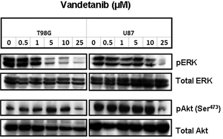
|
19622715 | |
| Growth inhibition assay | Cell viability |
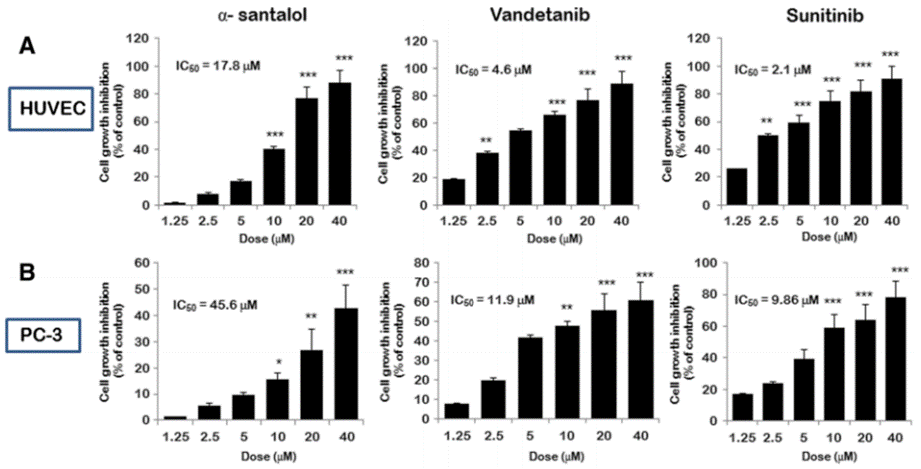
|
24261856 | |
| In Vivo | ||
| In Vivo | Vandetanib (2.5 mg/kg, i.v.), reverses a VEGF-induced hypotension by 63% but does not significantly affect a bFGF-induced hypotension. This compound (100 mg/kg) inhibits the tumor-induced blood vessel formation by 79%. It (12.5-100 mg/kg, orally) shows great tumor growth inhibition in human tumor xenografts including Calu-6, PC-3, MDA-MA-231, SKOV-3, SW620, A549, A431, B16-F10(AP3) and Lewis Lung, with little effects on body weight. In PC3wt xenografts, administration of this compound alone exerts paradoxical tumor growth stimulating effects. In PC3R xenografts, the low dose of this chemical (25 mg/kg) has no significant effect relative to control, whereas the high dose (50 mg/kg) significantly inhibits tumor growth compared with control. In contrast, the high-dose combination reveals a significant negative interaction between this compound 50 mg/kg and docetaxel 30 mg/kg in PC3R cells. In tumor-bearing mice, it suppresses phosphorylation of VEGFR2 and EGFR in tumor tissues, significantly decreases tumor vessel density, enhances tumor cell apoptosis, suppresses tumor growth, improves survival, reduces number of intrahepatic metastases, and up-regulates VEGF, TGF-alpha and EGF in tumor tissues. Treatment with this compound is not associated with serious adverse events, including ALT abnormality, bone marrow suppression or body weight loss. This chemical treatment of nude mice bearing palpable GEO colon cancer xenografts (which are sensitive to inhibition of EGFR signaling) induces dose-dependent tumor growth inhibition. | |
|---|---|---|
| 動物実験 | 動物モデル | Female athymic (nu/nu genotype) Swiss mice with PC-3, Calu-6, SKOV-3, and MDA-MB-231 tumors |
| 投与量 | 12.5 mg/kg/day, 25 mg/kg/day, 50 mg/kg/day, or 100 mg/kg/day | |
| 投与経路 | Oral administration | |
| NCT Number | Recruitment | Conditions | Sponsor/Collaborators | Start Date | Phases |
|---|---|---|---|---|---|
| NCT03291379 | Completed | Carcinoma Hepatocellular|Metastatic Colorectal Cancer |
Boston Scientific Corporation|Biocompatibles UK Ltd |
May 17 2017 | Early Phase 1 |
| NCT02495103 | Terminated | Renal Cell Carcinoma|Hereditary Leiomyomatosis|Renal Cell Cancer |
National Cancer Institute (NCI)|National Institutes of Health Clinical Center (CC) |
August 26 2015 | Phase 1|Phase 2 |
| NCT02530411 | Unknown status | Neoplasms |
Velindre NHS Trust|Cancer Research UK|AstraZeneca |
April 2015 | Phase 2 |
| NCT02268734 | Completed | Metastatic Sporadic Medullary Thyroid Cancer |
Fondazione IRCCS Istituto Nazionale dei Tumori Milano |
April 2014 | -- |
| NCT01876784 | Completed | Differentiated Thyroid Cancer |
Genzyme a Sanofi Company|Sanofi |
September 17 2013 | Phase 3 |
| NCT01661179 | Completed | Unresectable Locally Advanced or Metastatic Medullary Thyroid Carcinoma |
Genzyme a Sanofi Company|Sanofi |
November 2012 | Phase 1|Phase 2 |
|
化学情報
| 分子量 | 475.35 | 化学式 | C22H24BrFN4O2 |
| CAS No. | 443913-73-3 | SDF | Download Vandetanib (ZD6474) SDFをダウンロードする |
| Smiles | CN1CCC(CC1)COC2=C(C=C3C(=C2)N=CN=C3NC4=C(C=C(C=C4)Br)F)OC | ||
| 保管 | |||
|
In vitro |
DMSO : 60 mg/mL ( (126.22 mM); Warmed with 60℃ water bath; Ultrasonicated; 吸湿したDMSOは溶解度を減少させます。新しいDMSOをご使用ください。) Water : Insoluble Ethanol : Insoluble |
モル濃度計算器 |
|
in vivo Add solvents to the product individually and in order. |
投与溶液組成計算機 | |||||
実験計算
投与溶液組成計算機(クリア溶液)
ステップ1:実験データを入力してください。(実験操作によるロスを考慮し、動物数を1匹分多くして計算・調製することを推奨します)
mg/kg
g
μL
匹
ステップ2:投与溶媒の組成を入力してください。(ロット毎に適した溶解組成が異なる場合があります。詳細については弊社までお問い合わせください)
% DMSO
%
% Tween 80
% ddH2O
%DMSO
%
計算結果:
投与溶媒濃度: mg/ml;
DMSOストック溶液調製方法: mg 試薬を μL DMSOに溶解する(濃度 mg/mL, 注:濃度が当該ロットのDMSO溶解度を超える場合はご連絡ください。 )
投与溶媒調製方法:Take μL DMSOストック溶液に μL PEG300,を加え、完全溶解後μL Tween 80,を加えて完全溶解させた後 μL ddH2O,を加え完全に溶解させます。
投与溶媒調製方法:μL DMSOストック溶液に μL Corn oil,を加え、完全溶解。
注意:1.ストック溶液に沈殿、混濁などがないことをご確認ください;
2.順番通りに溶剤を加えてください。次のステップに進む前に溶液に沈殿、混濁などがないことを確認してから加えてください。ボルテックス、ソニケーション、水浴加熱など物理的な方法で溶解を早めることは可能です。
技術サポート
ストックの作り方、阻害剤の保管方法、細胞実験や動物実験の際に注意すべき点など、製品を取扱う時に問い合わせが多かった質問に対しては取扱説明書でお答えしています。
他に質問がある場合は、お気軽にお問い合わせください。
* 必須
納期 国内在庫品:受注日の翌日(15時までの受注分) *北海道、九州、沖縄への配送は受注日より2日以上 を要する場合あり 海外在庫品:受注後1〜2週間