
- 阻害剤
- 研究分野別
- PI3K/Akt/mTOR
- Epigenetics
- Methylation
- Immunology & Inflammation
- Protein Tyrosine Kinase
- Angiogenesis
- Apoptosis
- Autophagy
- ER stress & UPR
- JAK/STAT
- MAPK
- Cytoskeletal Signaling
- Cell Cycle
- TGF-beta/Smad
- 化合物ライブラリー
- 抗体
- 新製品
- お問い合わせ
Roscovitine (Seliciclib)
別名:CYC202, Seliciclib, R-roscovitine
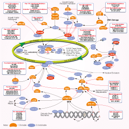
Roscovitineは、Cdc2、CDK2、およびCDK5に対する強力かつ選択的なCDK阻害剤であり、Cell CycleアッセイでのIC50はそれぞれ0.65 μM、0.7 μM、0.16 μMです。CDK4/6にはほとんど影響を示しません。フェーズ2。
CAS No. 186692-46-6
文献中Selleckの製品使用例(128)
製品安全説明書
現在のバッチを見る:
純度:
99.85%
99.85
Roscovitine (Seliciclib)関連製品
シグナル伝達経路
CDK阻害剤の選択性比較
Cell Data
| Cell Lines | Assay Type | Concentration | Incubation Time | 活性情報 | PMID |
|---|---|---|---|---|---|
| LP-1 | Apoptosis assay | 30 uM | 3 hrs | Induction of apoptosis in human LP-1 cells at 30 uM after 3 hrs using TUNEL staining by flow cytometry | 15958589 |
| LP-1 | Cytotoxicity assay | 20 to 30 uM | 24 hrs | Cytotoxicity against human LP-1 cells assessed as reduction of cell viability at 20 to 30 uM treated for 24 hrs followed by washout measured after total 72 hrs growth period alamar blue assay relative to control | 15958589 |
| LP-1 | Apoptosis assay | 30 uM | 1.5 hrs | Induction of apoptosis in human LP-1 cells assessed as reduction of RNA polymerase 2 phosphoserine 2 level at 30 uM after 1.5 hrs by immunoblotting | 15958589 |
| LP-1 | Apoptosis assay | 30 uM | 3 hrs | Induction of apoptosis in human LP-1 cells assessed as reduction of Mcl-1 protein level at 30 uM after 3 hrs by immunoblotting | 15958589 |
| LP-1 | Apoptosis assay | 30 uM | 3 to 5 hrs | Induction of apoptosis in human LP-1 cells assessed as increase in level of cleaved PARP at 30 uM after 3 to 5 hrs by immunoblotting | 15958589 |
| NCI-H929 | Apoptosis assay | 30 uM | 5 hrs | Induction of apoptosis in human NCI-H929 cells assessed as increase in level of cleaved PARP at 30 uM after 5 hrs by immunoblotting | 15958589 |
| NCI-H929 | Apoptosis assay | 30 uM | 1.5 hrs | Induction of apoptosis in human NCI-H929 cells assessed as fast slow migrating hyperphosphorylated RNA polymerase 2O form at 30 uM after 1.5 hrs by immunoblotting | 15958589 |
| RPM18226 | Apoptosis assay | 30 uM | 1.5 hrs | Induction of apoptosis in human RPM18226 cells assessed as reduction of RNA polymerase 2 phosphoserine 2 level at 30 uM after 1.5 hrs by immunoblotting | 15958589 |
| RPM18226 | Apoptosis assay | 30 uM | 3 hrs | Induction of apoptosis in human RPM18226 cells assessed as reduction of Mcl-1 protein level at 30 uM after 3 hrs by immunoblotting | 15958589 |
| RPM18226 | Apoptosis assay | 30 uM | 3 to 5 hrs | Induction of apoptosis in human RPM18226 cells assessed as increase in level of cleaved PARP at 30 uM after 3 to 5 hrs by immunoblotting | 15958589 |
| NCI-H929 | Apoptosis assay | 30 uM | 3 hrs | Induction of apoptosis in human NCI-H929 cells assessed as changes in XIAP protein level at 30 uM after 3 hrs by immunoblotting | 15958589 |
| NCI-H929 | Apoptosis assay | 30 uM | 3 hrs | Induction of apoptosis in human NCI-H929 cells assessed as changes in survivin protein level at 30 uM after 3 hrs by immunoblotting | 15958589 |
| RPM18226 | Apoptosis assay | 30 uM | 3 hrs | Induction of apoptosis in human RPM18226 cells at 30 uM after 3 hrs using TUNEL staining by flow cytometry | 15958589 |
| NCI-H929 | Apoptosis assay | 30 uM | 1.5 hrs | Induction of apoptosis in human NCI-H929 cells assessed as reduction of RNA polymerase 2 phosphoserine 2 level at 30 uM after 1.5 hrs by immunoblotting | 15958589 |
| NCI-H929 | Apoptosis assay | 30 uM | 1.5 hrs | Induction of apoptosis in human NCI-H929 cells assessed as dephosphorylation of pRb at S249/T252 at 30 uM after 1.5 hrs by immunoblotting | 15958589 |
| NCI-H929 | Cytotoxicity assay | 20 to 30 uM | 16 hrs | Cytotoxicity against human NCI-H929 cells assessed as reduction of cell viability at 20 to 30 uM treated for 16 hrs followed by washout measured after total 72 hrs growth period alamar blue assay relative to control | 15958589 |
| NCI-H929 | Apoptosis assay | 30 uM | 3 hrs | Induction of apoptosis in human NCI-H929 cells assessed as reduction of Mcl-1 protein level at 30 uM after 3 hrs by immunoblotting | 15958589 |
| NCI-H929 | Apoptosis assay | 30 uM | 3 hrs | Induction of apoptosis in human NCI-H929 cells assessed as changes in Bcl-2 protein level at 30 uM after 3 hrs by immunoblotting | 15958589 |
| NCI-H929 | Apoptosis assay | 30 uM | 3 hrs | Induction of apoptosis in human NCI-H929 cells at 30 uM after 3 hrs using TUNEL staining by flow cytometry | 15958589 |
| NCI-H929 | Apoptosis assay | 30 uM | 1.5 hrs | Induction of apoptosis in human NCI-H929 cells assessed as reduction of RNA polymerase 2 phosphoserine 5 level at 30 uM after 1.5 hrs by immunoblotting | 15958589 |
| NCI-H929 | Apoptosis assay | 30 uM | 1.5 hrs | Induction of apoptosis in human NCI-H929 cells assessed as reduction of Hdm2 level at 30 uM after 1.5 hrs by immunoblotting | 15958589 |
| NCI-H929 | Apoptosis assay | 30 uM | 1.5 hrs | Induction of apoptosis in human NCI-H929 cells assessed as increase of p53 accumulation at 30 uM after 1.5 hrs by immunoblotting | 15958589 |
| HCT116 | Function assay | 30 to 40 umol/L | 24 hrs | Inhibition of cyclin A in human HCT116 cells assessed as decrease in protein level at 30 to 40 umol/L after 24 hrs by immunoblotting analysis | 21080703 |
| HCT116 | Function assay | 30 to 40 umol/L | 24 hrs | Inhibition of cyclin B in human HCT116 cells assessed as decrease in protein level at 30 to 40 umol/L after 24 hrs by immunoblotting analysis | 21080703 |
| HCT116 | Function assay | 30 to 40 umol/L | 24 hrs | Inhibition of cyclin D1 in human HCT116 cells assessed as decrease in protein level at 30 to 40 umol/L after 24 hrs by immunoblotting analysis | 21080703 |
| HCT116 | Function assay | 30 to 40 umol/L | 24 hrs | Inhibition of CDK2 in human HCT116 cells assessed as decrease in protein level at 30 to 40 umol/L after 24 hrs by immunoblotting analysis | 21080703 |
| HT-29 | Function assay | 2.5 to 40 uM | 24 hrs | Inhibition of retinoblastoma protein in human HT-29 cells assessed as reduction of cyclin A level at 2.5 to 40 uM after 24 hrs by immunoblotting | 21417417 |
| MCF7 | Cell cycle assay | 80 uM | 24 hrs | Cell cycle arrest in human MCF7 cells assessed as reduction of actively replicating DNA level at 80 uM after 24 hrs using propidium iodide and BrdU staining by flow cytometry | 21417417 |
| MCF7 | Function assay | 20 uM | 24 hrs | Induction of p53-dependent transcriptional activity in human MCF7 cells assessed as increase of p21 WAF1 level at 20 uM after 24 hrs by immunofluorescence assay | 21417417 |
| RPMI8226 | Cell cycle assay | 80 uM | 24 hrs | Cell cycle arrest in human RPMI8226 cells assessed as reduction of actively replicating DNA level at 80 uM after 24 hrs using propidium iodide and BrdU staining by flow cytometry | 21417417 |
| A549 | Apoptosis assay | 2 uM | 48 hrs | Induction of apoptosis in human A549 cells assessed as DNA fragmentation at 2 uM after 48 hrs by agarose gel electrophoresis | 23623491 |
| BJ | Function assay | 10 uM | 10 days | Suppression of senescence in human BJ cells assessed as increase in cell number at 10 uM after 10 days by senescence reversal assay | 24681986 |
| BJ | Function assay | 10 uM | 10 days | Inhibition of ataxia telangiectasia-mutated in human BJ cells assessed as increase in cell number at 10 uM after 10 days by senescence reversal assay | 24681986 |
| MCF7 | Function assay | 10 uM | 10 mins | Sensitization of infrared-induced DNA damage in human MCF7 cells assessed as reduction in colony formation at 10 uM pretreated for 10 mins followed by irradiation for 4 hrs measured after 10 days by crystal violet staining analysis | 26851505 |
| MCF7 | Cell cycle assay | 24 hrs | Cell cycle arrest in human MCF7 cells assessed as accumulation at G2/M phase after 24 hrs using propidium iodide and BrdU staining by flow cytometry | 21417417 | |
| RPMI8226 | Cell cycle assay | 24 hrs | Cell cycle arrest in human RPMI8226 cells assessed as accumulation at G2/M phase after 24 hrs using propidium iodide and BrdU staining by flow cytometry | 21417417 | |
| MCF7 | Cell cycle assay | 24 hrs | Cell cycle arrest in human MCF7 cells assessed as decrease in S phase cell population after 24 hrs using propidium iodide and BrdU staining by flow cytometry | 21417417 | |
| MCF7 | Cell cycle assay | 24 hrs | Cell cycle arrest in human MCF7 cells assessed as accumulation at sub-G1 phase after 24 hrs using propidium iodide and BrdU staining by flow cytometry | 21417417 | |
| RPMI8226 | Cell cycle assay | 24 hrs | Cell cycle arrest in human RPMI8226 cells assessed as accumulation at sub-G1 phase after 24 hrs using propidium iodide and BrdU staining by flow cytometry | 21417417 | |
| RPMI8226 | Cell cycle assay | 24 hrs | Cell cycle arrest in human RPMI8226 cells assessed as decrease in S phase cell population after 24 hrs using propidium iodide and BrdU staining by flow cytometry | 21417417 | |
| Sf9 | Function assay | 10 mins | Inhibition of His-6-tagged recombinant human CDK2/cyclinE expressed in baculovirus-infected sf9 cells using histone H1 as substrate after 10 mins by liquid scintillation counting in presence of [gamma-32P]ATP, IC50 = 0.1 μM. | 24417566 | |
| NCI-SNU-1 | Growth Inhibition Assay | IC50=31.1059 μM | SANGER | ||
| NKM-1 | Growth Inhibition Assay | IC50=31.1397 μM | SANGER | ||
| SIG-M5 | Growth Inhibition Assay | IC50=31.6833 μM | SANGER | ||
| SK-N-FI | Growth Inhibition Assay | IC50=31.7535 μM | SANGER | ||
| LOUCY | Growth Inhibition Assay | IC50=32.1253 μM | SANGER | ||
| Calu-6 | Growth Inhibition Assay | IC50=32.4745 μM | SANGER | ||
| GOTO | Growth Inhibition Assay | IC50=32.9129 μM | SANGER | ||
| NCI-H526 | Growth Inhibition Assay | IC50=33.4936 μM | SANGER | ||
| RKO | Growth Inhibition Assay | IC50=33.5969 μM | SANGER | ||
| NCI-H64 | Growth Inhibition Assay | IC50=33.8597 μM | SANGER | ||
| LP-1 | Growth Inhibition Assay | IC50=33.8908 μM | SANGER | ||
| KGN | Growth Inhibition Assay | IC50=34.2524 μM | SANGER | ||
| NCI-H2141 | Growth Inhibition Assay | IC50=34.6533 μM | SANGER | ||
| TE-10 | Growth Inhibition Assay | IC50=34.9422 μM | SANGER | ||
| K5 | Growth Inhibition Assay | IC50=35.0861 μM | SANGER | ||
| IMR-5 | Growth Inhibition Assay | IC50=35.3139 μM | SANGER | ||
| TE-441-T | Growth Inhibition Assay | IC50=36.1148 μM | SANGER | ||
| TE-6 | Growth Inhibition Assay | IC50=36.3246 μM | SANGER | ||
| MOLT-4 | Growth Inhibition Assay | IC50=36.3276 μM | SANGER | ||
| COLO-684 | Growth Inhibition Assay | IC50=37.012 μM | SANGER | ||
| LU-139 | Growth Inhibition Assay | IC50=37.1856 μM | SANGER | ||
| OPM-2 | Growth Inhibition Assay | IC50=37.2949 μM | SANGER | ||
| ML-2 | Growth Inhibition Assay | IC50=37.6712 μM | SANGER | ||
| RS4-11 | Growth Inhibition Assay | IC50=37.7069 μM | SANGER | ||
| MONO-MAC-6 | Growth Inhibition Assay | IC50=38.2477 μM | SANGER | ||
| NCI-H345 | Growth Inhibition Assay | IC50=38.9106 μM | SANGER | ||
| NTERA-S-cl-D1 | Growth Inhibition Assay | IC50=39.5842 μM | SANGER | ||
| NCI-H1882 | Growth Inhibition Assay | IC50=40.5998 μM | SANGER | ||
| LC-1F | Growth Inhibition Assay | IC50=41.5705 μM | SANGER | ||
| HT | Growth Inhibition Assay | IC50=42.0028 μM | SANGER | ||
| MLMA | Growth Inhibition Assay | IC50=42.2787 μM | SANGER | ||
| DG-75 | Growth Inhibition Assay | IC50=42.6546 μM | SANGER | ||
| GI-ME-N | Growth Inhibition Assay | IC50=42.6671 μM | SANGER | ||
| MS-1 | Growth Inhibition Assay | IC50=42.893 μM | SANGER | ||
| CGTH-W-1 | Growth Inhibition Assay | IC50=44.9697 μM | SANGER | ||
| NCI-H209 | Growth Inhibition Assay | IC50=46.0115 μM | SANGER | ||
| LB2518-MEL | Growth Inhibition Assay | IC50=47.0448 μM | SANGER | ||
| DU-4475 | Growth Inhibition Assay | IC50=48.4937 μM | SANGER | ||
| LB2241-RCC | Growth Inhibition Assay | IC50=48.6202 μM | SANGER | ||
| LB771-HNC | Growth Inhibition Assay | IC50=48.9212 μM | SANGER | ||
| NCI-H82 | Growth Inhibition Assay | IC50=31.0135 μM | SANGER | ||
| NCI-H510A | Growth Inhibition Assay | IC50=30.0329 μM | SANGER | ||
| ES3 | Growth Inhibition Assay | IC50=29.9582 μM | SANGER | ||
| BB30-HNC | Growth Inhibition Assay | IC50=29.9483 μM | SANGER | ||
| KM12 | Growth Inhibition Assay | IC50=29.6239 μM | SANGER | ||
| GI-1 | Growth Inhibition Assay | IC50=29.0113 μM | SANGER | ||
| NOS-1 | Growth Inhibition Assay | IC50=28.9733 μM | SANGER | ||
| TE-8 | Growth Inhibition Assay | IC50=28.908 μM | SANGER | ||
| TE-9 | Growth Inhibition Assay | IC50=28.7969 μM | SANGER | ||
| HL-60 | Growth Inhibition Assay | IC50=27.9869 μM | SANGER | ||
| QIMR-WIL | Growth Inhibition Assay | IC50=27.9144 μM | SANGER | ||
| KARPAS-299 | Growth Inhibition Assay | IC50=26.8646 μM | SANGER | ||
| KURAMOCHI | Growth Inhibition Assay | IC50=26.8082 μM | SANGER | ||
| BL-41 | Growth Inhibition Assay | IC50=25.9597 μM | SANGER | ||
| NCI-H2126 | Growth Inhibition Assay | IC50=25.6529 μM | SANGER | ||
| HOP-62 | Growth Inhibition Assay | IC50=25.4425 μM | SANGER | ||
| IST-SL2 | Growth Inhibition Assay | IC50=24.5343 μM | SANGER | ||
| HH | Growth Inhibition Assay | IC50=24.3819 μM | SANGER | ||
| LS-513 | Growth Inhibition Assay | IC50=23.5179 μM | SANGER | ||
| EB-3 | Growth Inhibition Assay | IC50=23.1831 μM | SANGER | ||
| ACN | Growth Inhibition Assay | IC50=21.3389 μM | SANGER | ||
| NOMO-1 | Growth Inhibition Assay | IC50=21.2008 μM | SANGER | ||
| ES8 | Growth Inhibition Assay | IC50=21.06 μM | SANGER | ||
| CESS | Growth Inhibition Assay | IC50=20.8549 μM | SANGER | ||
| BL-70 | Growth Inhibition Assay | IC50=20.3274 μM | SANGER | ||
| MHH-PREB-1 | Growth Inhibition Assay | IC50=20.0356 μM | SANGER | ||
| BC-1 | Growth Inhibition Assay | IC50=19.1198 μM | SANGER | ||
| LC4-1 | Growth Inhibition Assay | IC50=18.8734 μM | SANGER | ||
| COLO-320-HSR | Growth Inhibition Assay | IC50=18.7688 μM | SANGER | ||
| A101D | Growth Inhibition Assay | IC50=18.3208 μM | SANGER | ||
| BC-3 | Growth Inhibition Assay | IC50=18.0305 μM | SANGER | ||
| TGW | Growth Inhibition Assay | IC50=17.8124 μM | SANGER | ||
| JAR | Growth Inhibition Assay | IC50=17.0152 μM | SANGER | ||
| HD-MY-Z | Growth Inhibition Assay | IC50=16.8246 μM | SANGER | ||
| NCI-H1304 | Growth Inhibition Assay | IC50=16.3601 μM | SANGER | ||
| OS-RC-2 | Growth Inhibition Assay | IC50=15.8382 μM | SANGER | ||
| OCI-AML2 | Growth Inhibition Assay | IC50=15.6482 μM | SANGER | ||
| HCC1599 | Growth Inhibition Assay | IC50=14.5975 μM | SANGER | ||
| SCC-3 | Growth Inhibition Assay | IC50=14.2956 μM | SANGER | ||
| RPMI-6666 | Growth Inhibition Assay | IC50=13.9121 μM | SANGER | ||
| MEG-01 | Growth Inhibition Assay | IC50=13.8379 μM | SANGER | ||
| Raji | Growth Inhibition Assay | IC50=13.7894 μM | SANGER | ||
| RPMI-8402 | Growth Inhibition Assay | IC50=13.6262 μM | SANGER | ||
| GCIY | Growth Inhibition Assay | IC50=12.8613 μM | SANGER | ||
| 697 | Growth Inhibition Assay | IC50=12.6007 μM | SANGER | ||
| D-247MG | Growth Inhibition Assay | IC50=12.3516 μM | SANGER | ||
| NB1 | Growth Inhibition Assay | IC50=12.3308 μM | SANGER | ||
| COR-L279 | Growth Inhibition Assay | IC50=12.2907 μM | SANGER | ||
| LB831-BLC | Growth Inhibition Assay | IC50=11.5624 μM | SANGER | ||
| ST486 | Growth Inhibition Assay | IC50=10.351 μM | SANGER | ||
| SK-UT-1 | Growth Inhibition Assay | IC50=10.35 μM | SANGER | ||
| BB65-RCC | Growth Inhibition Assay | IC50=9.97495 μM | SANGER | ||
| KARPAS-422 | Growth Inhibition Assay | IC50=9.96336 μM | SANGER | ||
| Becker | Growth Inhibition Assay | IC50=9.46082 μM | SANGER | ||
| KS-1 | Growth Inhibition Assay | IC50=9.45785 μM | SANGER | ||
| JiyoyeP-2003 | Growth Inhibition Assay | IC50=8.50264 μM | SANGER | ||
| NCCIT | Growth Inhibition Assay | IC50=7.55482 μM | SANGER | ||
| MRK-nu-1 | Growth Inhibition Assay | IC50=7.12969 μM | SANGER | ||
| A3-KAW | Growth Inhibition Assay | IC50=5.76116 μM | SANGER | ||
| SK-N-MC | qHTS assay | qHTS of pediatric cancer cell lines to identify multiple opportunities for drug repurposing: Primary screen for SK-N-MC cells | 15958589 | ||
| SK-N-MC | qHTS assay | qHTS of pediatric cancer cell lines to identify multiple opportunities for drug repurposing: Primary screen for SK-N-MC cells | 21080703 | ||
| Caco2 | Cell cycle assay | Cell cycle arrest in human Caco2 cells assessed as accumulation at G1/S phase by Hoechst staining based fluorescence assay | 28214231 | ||
| HaCaT | Cell cycle assay | Cell cycle arrest in human HaCaT cells assessed as accumulation at G1/S phase by Hoechst staining based fluorescence assay | 28214231 | ||
| HuH7 | Cell cycle assay | Cell cycle arrest in human HuH7 cells assessed as accumulation at G1/S phase by Hoechst staining based fluorescence assay | 28214231 | ||
| PC3 | Cell cycle assay | Cell cycle arrest in human PC3 cells assessed as accumulation at G2/M phase by Hoechst staining based fluorescence assay | 28214231 | ||
| MDA-MB-231 | Cell cycle assay | Cell cycle arrest in human MDA-MB-231 cells assessed as accumulation at G1/S phase by Hoechst staining based fluorescence assay | 28214231 | ||
| HCT116 | Cell cycle assay | Cell cycle arrest in human HCT116 cells assessed as accumulation at G1/S phase by Hoechst staining based fluorescence assay | 28214231 | ||
| SK-N-MC | qHTS assay | qHTS of pediatric cancer cell lines to identify multiple opportunities for drug repurposing: Primary screen for SK-N-MC cells | 28557430 | ||
| A673 | qHTS assay | qHTS of pediatric cancer cell lines to identify multiple opportunities for drug repurposing: Primary screen for A673 cells | 29435139 | ||
| DAOY | qHTS assay | qHTS of pediatric cancer cell lines to identify multiple opportunities for drug repurposing: Primary screen for DAOY cells | 29435139 | ||
| BT-37 | qHTS assay | qHTS of pediatric cancer cell lines to identify multiple opportunities for drug repurposing: Primary screen for BT-37 cells | 29435139 | ||
| SJ-GBM2 | qHTS assay | qHTS of pediatric cancer cell lines to identify multiple opportunities for drug repurposing: Primary screen for SJ-GBM2 cells | 29435139 | ||
| LAN-5 | qHTS assay | qHTS of pediatric cancer cell lines to identify multiple opportunities for drug repurposing: Primary screen for LAN-5 cells | 29435139 | ||
| SK-N-MC | qHTS assay | qHTS of pediatric cancer cell lines to identify multiple opportunities for drug repurposing: Primary screen for SK-N-MC cells | 30199702 | ||
| 他の多くの細胞株試験データをご覧になる場合はこちらをクリックして下さい | |||||
生物活性
| 製品説明 | Roscovitineは、Cdc2、CDK2、およびCDK5に対する強力かつ選択的なCDK阻害剤であり、Cell CycleアッセイでのIC50はそれぞれ0.65 μM、0.7 μM、0.16 μMです。CDK4/6にはほとんど影響を示しません。フェーズ2。 | ||||||||||
|---|---|---|---|---|---|---|---|---|---|---|---|
| Targets |
|
| In Vitro | ||||
| In vitro |
Roscovitine displays high efficiency and high selectivity towards some cyclin-dependent kinases with IC50 of 0.65, 0.7, 0.7 and 0.16 μM for cdc2/cyclin B, cdk2/cyclin A, cdk2/cyclin E and cdk5/p53, respectively. This compound reversibly inhibits the prophaselmetaphase transition in the micromolar range of starfish oocytes and sea urchin embryos, inhibits in vitro M-phase-promoting factor activity and in vitro DNA synthesis in Xenopus egg extracts, and suppresses the proliferation of mammalian cell lines with an average IC50 of 16 μM. In mesangial cells, it results in a dose-dependent reduction of CDK2 activity that at concentrations of 7.5, 12.5 and 25 mM, this chemical causes a 25, 50% and 100% decrease in CDK2 activity, respectively. A recent study shows that this compound inhibits cdk5 kinase activity, cell proliferation, multicellular development, and cdk5 nuclear translocation in Dictyostelium discoideum, without affecting the expression of cdk5 protein during axenic growth. |
|||
|---|---|---|---|---|
| Kinase Assay | Enzymes | |||
| [API调用失败: invalid chat.event: ping, {'event': 'ping', 'data': '{"timestamp_ms":"1763706911366"}'}] | ||||
| 細胞実験 | 細胞株 | Leukemia, non-small cell lung cancer, colon cancer, central nervous system cancer, melanoma, ovarian cancer, renal cancer, prostate cancer, breast cancer | ||
| 濃度 | 0.01 - 100 μM | |||
| 反応時間 | 48 hours | |||
| 実験の流れ | 60 human tumour cell lines comprising nine tumor types are cultured for 24 hours prior to a 48-hour continuous exposure to 0.01-100 μM roscovitine. A sulforhodaminine B protein assay is used to estimate the cytotoxicity. |
|||
| 実験結果図 | Methods | Biomarkers | 結果図 | PMID |
| Western blot | pT231-tau / pS202-tau / tau p-Rb / p-CDK2 / CDK2 / Cyclin D1 / Cyclin A2 / ERα / ERβ/ AIB1 / PELP1 |
|
30915013 | |
| Immunofluorescence | CDK1 / Smek2 / FUBP1 / Cdc20 E2F1 / FASN / Bmi1 / Cyclin D2 / CDK2 / CDK4 |
|
24534090 | |
| Growth inhibition assay | Cell viability |
|
29996940 | |
| In Vivo | ||
| In Vivo |
Roscovitine, at a dose of 50 mg/kg, significantly inhibits growth of The Ewing's sarcoma family of tumors (ESFT) xenografts. This compound enhances the antitumor effect of doxorubicin without increased toxicity with a mechanism that involves cell cycle arrest rather than apoptosis in nude mice bearing established MCF7 xenografts. |
|
|---|---|---|
| 動物実験 | 動物モデル | A4573 cells are injected s.c. into the right posterior flank of CD1 nu/nu mice. |
| 投与量 | ≤50 mg/kg | |
| 投与経路 | Administered via i.p. | |
| NCT Number | Recruitment | Conditions | Sponsor/Collaborators | Start Date | Phases |
|---|---|---|---|---|---|
| NCT02649751 | Terminated | Cystic Fibrosis |
University Hospital Brest|ManRos Therapeutics|Cyclacel Pharmaceuticals Inc. |
February 22 2016 | Phase 2 |
|
化学情報
| 分子量 | 354.45 | 化学式 | C19H26N6O |
| CAS No. | 186692-46-6 | SDF | Download Roscovitine (Seliciclib) SDFをダウンロードする |
| Smiles | CCC(CO)NC1=NC(=C2C(=N1)N(C=N2)C(C)C)NCC3=CC=CC=C3 | ||
| 保管 | |||
|
In vitro |
DMSO : 71 mg/mL ( (200.31 mM); 吸湿したDMSOは溶解度を減少させます。新しいDMSOをご使用ください。) Ethanol : 71 mg/mL Water : Insoluble |
モル濃度計算器 |
|
in vivo Add solvents to the product individually and in order. |
投与溶液組成計算機 | |||||
実験計算
投与溶液組成計算機(クリア溶液)
ステップ1:実験データを入力してください。(実験操作によるロスを考慮し、動物数を1匹分多くして計算・調製することを推奨します)
mg/kg
g
μL
匹
ステップ2:投与溶媒の組成を入力してください。(ロット毎に適した溶解組成が異なる場合があります。詳細については弊社までお問い合わせください)
% DMSO
%
% Tween 80
% ddH2O
%DMSO
%
計算結果:
投与溶媒濃度: mg/ml;
DMSOストック溶液調製方法: mg 試薬を μL DMSOに溶解する(濃度 mg/mL, 注:濃度が当該ロットのDMSO溶解度を超える場合はご連絡ください。 )
投与溶媒調製方法:Take μL DMSOストック溶液に μL PEG300,を加え、完全溶解後μL Tween 80,を加えて完全溶解させた後 μL ddH2O,を加え完全に溶解させます。
投与溶媒調製方法:μL DMSOストック溶液に μL Corn oil,を加え、完全溶解。
注意:1.ストック溶液に沈殿、混濁などがないことをご確認ください;
2.順番通りに溶剤を加えてください。次のステップに進む前に溶液に沈殿、混濁などがないことを確認してから加えてください。ボルテックス、ソニケーション、水浴加熱など物理的な方法で溶解を早めることは可能です。
技術サポート
ストックの作り方、阻害剤の保管方法、細胞実験や動物実験の際に注意すべき点など、製品を取扱う時に問い合わせが多かった質問に対しては取扱説明書でお答えしています。
他に質問がある場合は、お気軽にお問い合わせください。
* 必須
よくある質問(FAQ)
質問1:
How can I reconstitute it for in vivo studies?
回答
It in 1% DMSO+10% Tween 80+20% N-N-dimethylacetamide+PEG 400 is a clear solution which is okay for injection. And this compound in 1% DMSO+30% polyethylene glycol+1% Tween 80 at 30mg/ml is a suspension, which is fine for oral gavage.
納期 国内在庫品:受注日の翌日(15時までの受注分) *北海道、九州、沖縄への配送は受注日より2日以上 を要する場合あり 海外在庫品:受注後1〜2週間