
- 阻害剤
- 研究分野別
- PI3K/Akt/mTOR
- Epigenetics
- Methylation
- Immunology & Inflammation
- Protein Tyrosine Kinase
- Angiogenesis
- Apoptosis
- Autophagy
- ER stress & UPR
- JAK/STAT
- MAPK
- Cytoskeletal Signaling
- Cell Cycle
- TGF-beta/Smad
- 化合物ライブラリー
- 抗体
- 新製品
- お問い合わせ
3-Methyladenine (3-MA)
別名:NSC 66389
3-Methyladenine (3-MA)は、HeLa細胞におけるVps34およびPI3Kγに対する選択的PI3K阻害剤であり、IC50はそれぞれ25 μMと60 μMです。クラスI PI3Kを一貫して阻害する一方、クラスIII PI3Kの抑制は一時的であり、autophagosome形成も阻害します。溶液は不安定なため、使用直前に調製する必要があります。

CAS No. 5142-23-4
文献中Selleckの製品使用例(956)
製品安全説明書
現在のバッチを見る:
純度:
99.97%
99.97
3-Methyladenine (3-MA)と併用されることが多い化合物
It and Necrostatin-1 inhibit cell death of bone marrow macrophages (BMDM) induced by LPS/zVAD and PolyI: C/zVAD.
This compound and Z-VAD-FMK combination confirm vital role of programmed cell death in pristimerin-mediated anti-cancer actions.
This compound and Ferrostatin-1 use abolishes acrylamide (ACR)-induced cell death in chondrocytes.
3-Methyladenine (3-MA)関連製品
シグナル伝達経路
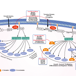
PI3K阻害剤の選択性比較
Cell Data
| Cell Lines | Assay Type | Concentration | Incubation Time | 活性情報 | PMID |
|---|---|---|---|---|---|
| SMMC-7721 | Apoptosis Assay | 5mM | 24h | attenuates TNF-α protection against serum starvation-mediated apoptosis | 24066693 |
| HO8910 | Apoptosis Assay | 10mM | 12h | enhances B19-induced apoptosi | 23983610 |
| MCF7 | Function Assay | 5mM | 24h | increases CuO induced cell death | 23962629 |
| HONE-1 | Function Assay | 5mM | 1h | represses 6r-mediated ROS production | 23892358 |
| HeLa | Function Assay | 10mM | 2h | suppresses LC3 II expressison | 23864738 |
| HepG2 | Function Assay | 10mM | 24h | inhibits siTIGAR- and HBSS-induced autophagy | 23817040 |
| SH-SY5Y | Function Assay | 1mM | 24h | inhibits the autophagy induced by TOCP | 23743148 |
| SH-SY5Y | Apoptosis Assay | 5mM | 1h | abolishes celastrol neuroprotective effect | 23619395 |
| PC12 | Function Assay | 10mM | 24h | inhibits chymotrypsin-like proteasomal activity. | 23603979 |
| OV2008 | Apoptosis Assay | 5mM | 24h | converts FTY720 with CDDP into an additive effect towards killing ovarian cancer cells | 23592281 |
| A2780 | Apoptosis Assay | 5mM | 24h | converts FTY720 with CDDP into an additive effect towards killing ovarian cancer cells | 23592281 |
| 1321N1 | Cytotoxicity Assay | 5mM | 24h | protects cell against PCN-induced toxicity | 23525265 |
| Saos-2 | Apoptosis Assay | 1mM | 96h | increases cell death induced by PCX | 23563171 |
| SH-SY5Y | Cytotoxicity Assay | 5mM | 24h | increases PCN toxicity | 23525265 |
| HT-29 | Function Assay | 1mM | 48/96h | inhibits AMPK induces autophagic cell death | 23508272 |
| OR6 | Function Assay | 10mM | 72h | suppresses HCV replication and formation of autophagosomes | 23395875 |
| Hela | Function Assay | 5mM | 24h | inhibits starvation-induced autophagy | 23395679 |
| MCF-7 | Function Assay | 5mM | 24h | inhibits starvation-induced autophagy | 23395679 |
| HUVECs | Function Assay | 3mM | 24h | blocks the protective effect of resveratrol by inhibiting autophagy | 23358928 |
| T24 | Function Assay | 10mM | 1h | reduces the cleavage of LC3 after baicalin treatment | 23354080 |
| U251MG | Function Assay | 3mM | 1h | suppresses LC3-II protein expression | 23338618 |
| GTL-16 | Apoptosis Assay | 5mM | 24h | reduces cell viability as compared to cells treated with MET inhibitors | 23313490 |
| T-47D | Function Assay | 10mM | 2h | inhibits autophagy process and increases rapamycin induced apoptosis | 23300026 |
| PaCa44 | Apoptosis Assay | 2.5mM | 1h | reduces genipin-mediated apoptosis | 23124112 |
| MDA-MB231 | Function Assay | 5mM | 1h | increases resveratrol-mediated caspase activation and cell death | 23088850 |
| SK-HEP-1 | Apoptosis Assay | 10mM | 1h | protects against autophagy and induces apoptosis in bufalin-treated cells | 22858649 |
| HeLa | Apoptosis Assay | 10mM | 2h | decreases cell viability co-treatment with PEI | 23000135 |
| HepG2 | Apoptosis Assay | 3mM | 5h | reduces cell apoptosis induced by QDs | 22836595 |
| MCF-7 | Function Assay | 2mM | 24h | inhibits autophagy induced by TM | 24970676 |
| MCF-7 | Function Assay | 2mM | 48h | promotes TM-induced cell death | 24970676 |
| MDA-MB-231 | Function Assay | 2mM | 24h | inhibits autophagy induced by TM | 24970676 |
| MDA-MB-231 | Function Assay | 2mM | 48h | promotes TM-induced cell death | 24970676 |
| PANC-1 | Apoptosis Assay | 1mM | 48h | enhances bortezomib-induced cell viability loss | 24842158 |
| MDA-MB 231 | Apoptosis Assay | 5mM | 0.5h | modulates Tocomin® induced apoptosis | 24830781 |
| Microglia | Apoptosis Assay | 5mM | 24h | decreases hypoxia-induced cell death | 24818601 |
| HepG2 | Function Assay | 5mM | 4h | increases cellular levels of HL | 24713587 |
| A2780cp | Apoptosis Assay | 2.5mM | 1h | enhances cisplatin-induced cell death | 24817946 |
| U2OS | Growth Inhibition Assay | 10mM | 24h | intensifies the growth inhibition induced by Dox | 24639013 |
| HCT116 | Apoptosis Assay | 5mM | 24h | enhances apigenin-induced cell death | 24626522 |
| PC-3 | Apoptosis Assay | 2mM | 2h | increases ORI-induced cell death | 22745580 |
| MCF-7 | Apoptosis Assay | 0.1mM | 6h | enhances sirtinol-induced apoptosis | 22751989 |
| U251 | Apoptosis Assay | 5mM | 24h | increases S1-induced cell death | 22579788 |
| HeLa | Apoptosis Assay | 5mM | 24h | induces caspase-dependent cell death | 22545128 |
| pDCs | Function Assay | 10mM | 0.5h | reduces the induction of IFN-α by ssRNA40 | 22396599 |
| A549 | Function Assay | 0.1mM | 24h | suppresses SU11274-induced cell death | 22466960 |
| BGC-823 | Function Assay | 5mM | 2h | inhibits the rate of autophagic cells | 22322152 |
| U937 | Function Assay | 2mM | 12h | decreases the autophagy ratio | 22155150 |
| Marc-145 | Function Assay | 5mM | 12/24/36h | reduces the PRRSV titers and the protein expression | 22119900 |
| HBx | Apoptosis Assay | 10mM | 48h | increases cell death | 22020078 |
| MCF-7 | Function Assay | 10mM | 48h | blocks autophagy induced by bortezomib | 21931937 |
| RMPI8226 | Function Assay | 5mM | 1h | suppresses the level of autophagy under nutrient depletion | 21915620 |
| PC12/TetOn | Function Assay | 0.1/1mM | 18h | leads to α-syn(WT) accumulation, toxicity, and oligomer formation | 21906659 |
| HeLa | Cytotoxicity Assay | 2mM | 24h | inhibites the cytotoxicity of silibinin to HeLa cells. | 21875385 |
| Jurkat | Function Assay | 10mM | 1h | decreases the expression of LC3-II and the formation of autophagosomes | 21864037 |
| K562 | Function Assay | 10mM | 1h | decreases the expression of LC3-II and the formation of autophagosomes | 21864037 |
| Hep3B | Apoptosis Assay | 5mM | 24h | attenuates TNF-α protection against serum starvation-mediated apoptosis | 24066693 |
| H460 | Function Assay | 10mM | 4h | increases cisplatin-induced cell death | 24173208 |
| A549 | Function Assay | 10mM | 4h | inhibits autophagy induced by irradiation | 24142735 |
| H1299 | Function Assay | 10mM | 4h | increases cisplatin-induced cell death | 24173208 |
| WiDr | Function Assay | 10mM | 1h | inhibits PCBL-induced LC3 II expression | 24190489 |
| LoVo | Apoptosis Assay | 5mM | 48h | enhances DCA-induced apoptosis. | 24201812 |
| HepG2 E47 | Function Assay | 2.5mM | 48h | increases the toxicity of AA, BSO, and CCl4 | 24273738 |
| RKO | Function Assay | 2mM | 1h | enhances cell death by geldanamycin | 24291777 |
| Hep3B | Apoptosis Assay | 2mM | 12h | inhibits AZD8055-induced cell death | 24297300 |
| ACHN-5968 | Apoptosis Assay | 5mM | 3h | enhances paclitaxel-mediated apoptosis | 24305604 |
| Huh7 | Apoptosis Assay | 2mM | 12h | inhibits AZD8055-induced cell death | 24297300 |
| UOK257 | Apoptosis Assay | 5mM | 3h | enhances paclitaxel-mediated apoptosis | 24305604 |
| ECSCs | Apoptosis Assay | 10mM | 4h | decreases rapamycin-treated apoptosis | 24319109 |
| MCF-7 | Function Assay | 10mM | 24h | inhibits the autophagy induced by chemotherapy drugs | 24315578 |
| SGC-7901 | Apoptosis Assay | 2mM | 1h | increases CA-4 induced apoptosis | 24321340 |
| SMMC-7721 | Apoptosis Assay | 2mM | 1h | increases CA-4 induced apoptosis | 24321340 |
| T24 | Apoptosis Assay | 5mM | 1.5h | potentiates celecoxib-induced apoptosis | 24349176 |
| NTUB1 | Apoptosis Assay | 5mM | 1.5h | potentiates celecoxib-induced apoptosis | 24349176 |
| MG-63 | Apoptosis Assay | 10mM | 12h | enhances DP-induced apoptosis | 24358301 |
| MG-63 | Apoptosis Assay | 0.5/1mM | 32h | enhances salinomycin-induced cell apoptosis | 24358342 |
| MG-63 | Function Assay | 0.5/1mM | 48h | induces salinomycin-induced cell viability loss | 24358342 |
| U2OS | Function Assay | 0.5/1mM | 48h | induces salinomycin-induced cell viability loss | 24358342 |
| HGC-27 | Function Assay | 10mM | 1h | inhibits the cell viability loss by RAD001 or MK-2206 | 24416349 |
| HCT116 | Apoptosis Assay | 5mM | 24h | enhances the apoptosis induced by apigenin | 24626522 |
| A549 | Apoptosis Assay | 10mM | 48h | accelerates the reduction of cell viability induced by PTX | 24626722 |
| Saos-2 | Apoptosis Assay | 10mM | 24h | intensifies the growth inhibition of the U2OS cells induced by Dox | 24639013 |
| U2OS | Apoptosis Assay | 10mM | 24h | intensifies the growth inhibition of the U2OS cells induced by Dox | 24639013 |
| HepG2 | Function Assay | 5mM | 4h | increases HL release | 24713587 |
| A549 | Apoptosis Assay | 5mM | 48h | decreases the percentage of cell death and expression levels of caspase-3, Beclin-1 and LC3-II | 24706303 |
| A2780cp | Apoptosis Assay | 2.5mM | 1h | enhances cisplatin-induced cell death | 24817946 |
| Microglia | Apoptosis Assay | 5mM | 24h | decreases hypoxia-induced cell death | 24818601 |
| HT-29 | Apoptosis Assay | 1mM | 48h | enhances bortezomib-induced cell viability loss | 24842158 |
| MDR | Apoptosis Assay | 10mM | 6h | strengthens the power of anticancer drugs | 25019701 |
| H157 | Function Assay | 5mM | 2h | suppresses SPC induced accumulation of LC3-II | 25285628 |
| A549 | Function Assay | 5mM | 2h | suppresses SPC induced accumulation of LC3-II | 25285628 |
| A2780cp | Growth Inhibition Assay | 1mM | 1h | increases cisplatin-induced cell death | 25322694 |
| NBL-W-S | Apoptosis Assay | 1mM | 6h | increases cell apoptosis induced by GANT-61 | 25323222 |
| NBL-W-S | Growth Inhibition Assay | 1mM | 6h | enhances GANT-61 toxicity | 25323222 |
| A549 | Apoptosis Assay | 5mM | 2h | inhibits BDMC-induced apoptotic cell death | 25716561 |
| 95D | Apoptosis Assay | 5mM | 2h | inhibits BDMC-induced apoptotic cell death | 25716561 |
| A549 | Growth Inhibition Assay | 3mM | 2h | reduces growth inhibitory effect of BDMC | 25716561 |
| 95D | Growth Inhibition Assay | 3mM | 2h | reduces growth inhibitory effect of BDMC | 25716561 |
| Nara-H | Growth Inhibition Assay | 5mM | 48h | enhances temsirolimusmediated suppression of Nara-H cell proliferation | 21805033 |
| HUVECs | Function Assay | 10mM | 0.5h | decreases the AGE-BSAinduced autophagy leve | 21468592 |
| HepG2 | Apoptosis Assay | 2mM | 1h | enhances radiation-induced cell death | 21453691 |
| U-2 OS | qHTS assay | qHTS of pediatric cancer cell lines to identify multiple opportunities for drug repurposing: Primary screen for U-2 OS cells | 29435139 | ||
| A673 | qHTS assay | qHTS of pediatric cancer cell lines to identify multiple opportunities for drug repurposing: Primary screen for A673 cells | 29435139 | ||
| DAOY | qHTS assay | qHTS of pediatric cancer cell lines to identify multiple opportunities for drug repurposing: Primary screen for DAOY cells | 29435139 | ||
| Saos-2 | qHTS assay | qHTS of pediatric cancer cell lines to identify multiple opportunities for drug repurposing: Primary screen for Saos-2 cells | 29435139 | ||
| BT-37 | qHTS assay | qHTS of pediatric cancer cell lines to identify multiple opportunities for drug repurposing: Primary screen for BT-37 cells | 29435139 | ||
| RD | qHTS assay | qHTS of pediatric cancer cell lines to identify multiple opportunities for drug repurposing: Primary screen for RD cells | 29435139 | ||
| SK-N-SH | qHTS assay | qHTS of pediatric cancer cell lines to identify multiple opportunities for drug repurposing: Primary screen for SK-N-SH cells | 29435139 | ||
| BT-12 | qHTS assay | qHTS of pediatric cancer cell lines to identify multiple opportunities for drug repurposing: Primary screen for BT-12 cells | 29435139 | ||
| MG 63 (6-TG R) | qHTS assay | qHTS of pediatric cancer cell lines to identify multiple opportunities for drug repurposing: Primary screen for MG 63 (6-TG R) cells | 29435139 | ||
| NB1643 | qHTS assay | qHTS of pediatric cancer cell lines to identify multiple opportunities for drug repurposing: Primary screen for NB1643 cells | 29435139 | ||
| OHS-50 | qHTS assay | qHTS of pediatric cancer cell lines to identify multiple opportunities for drug repurposing: Primary screen for OHS-50 cells | 29435139 | ||
| Rh41 | qHTS assay | qHTS of pediatric cancer cell lines to identify multiple opportunities for drug repurposing: Primary screen for Rh41 cells | 29435139 | ||
| Rh30 | qHTS assay | qHTS of pediatric cancer cell lines to identify multiple opportunities for drug repurposing: Primary screen for Rh30 cells | 29435139 | ||
| SJ-GBM2 | qHTS assay | qHTS of pediatric cancer cell lines to identify multiple opportunities for drug repurposing: Primary screen for SJ-GBM2 cells | 29435139 | ||
| SK-N-MC | qHTS assay | qHTS of pediatric cancer cell lines to identify multiple opportunities for drug repurposing: Primary screen for SK-N-MC cells | 29435139 | ||
| NB-EBc1 | qHTS assay | qHTS of pediatric cancer cell lines to identify multiple opportunities for drug repurposing: Primary screen for NB-EBc1 cells | 29435139 | ||
| LAN-5 | qHTS assay | qHTS of pediatric cancer cell lines to identify multiple opportunities for drug repurposing: Primary screen for LAN-5 cells | 29435139 | ||
| Rh18 | qHTS assay | qHTS of pediatric cancer cell lines to identify multiple opportunities for drug repurposing: Primary screen for Rh18 cells | 29435139 | ||
| 他の多くの細胞株試験データをご覧になる場合はこちらをクリックして下さい | |||||
生物活性
| 製品説明 | 3-Methyladenine (3-MA)は、HeLa細胞におけるVps34およびPI3Kγに対する選択的PI3K阻害剤であり、IC50はそれぞれ25 μMと60 μMです。クラスI PI3Kを一貫して阻害する一方、クラスIII PI3Kの抑制は一時的であり、autophagosome形成も阻害します。溶液は不安定なため、使用直前に調製する必要があります。 | ||||||
|---|---|---|---|---|---|---|---|
| Targets |
|
| In Vitro | ||||
| In vitro |
The slight preference for Vps34 prevention by 3-Methyladenine (3-MA) probably arises from a hydrophobic ring specific to Vps34, which encircles the 3-methyl group of this compound. It has been reported to cause cancer cell death under both normal and starvation conditions, and could also suppress cell migration and invasion independently of its ability to inhibit autophagy, implying that it possesses functions other than autophagy suppression. This compound elicits caspase-dependent cell death that is independent of autophagy inhibition. Treatment with 5 mM of it reduces the percentage of glucose-starved HeLa cells displaying GFP-LC3 puncta to 23%. The levels of LC3-I are increasing and the levels of LC3-II are decreasing between 12 and 48 hours in cells that are treated with 3-MA. Conversion of LC3-I to LC3-II is suppressed by the compound. Treatment of HeLa cells with it at 2.5 mM or 5 mM for one day does not affect cell viability, whereas treatment with 10 mM for one day causes a 25.0% decrease in cell viability. Treatment of cells with 2.5, 5 or 10 mM for two days causes 11.5%, 38.0% and 79.4% decrease in viability, respectively. It decreases cell viability in a time- and dose-dependent manner, and significantly shortens the duration of nocodazole-induced-prometaphase arrest. Suppression of autophagy by 3-MA inhibits SU11274-induced cell death. Prolonged treatment with it (up to 9 hours) induces significant LC3 I to II conversion in wild type MEFs. Prolonged treatment with 3-MA, but not wortmannin, markedly increases GFP-LC3 punctuation/aggregation. Its induced LC3 conversion and free GFP liberation are ATG7-dependent. Treatment with it leads to evident increase of p62 protein level. The compound increases the p62 level even in Atg5−/− MEFs as well as in cells with DOX-mediated deletion of ATG5. It inhibits class I and class III PI3K in different temporal patterns. Its induced LC3 I to LC3 II conversion is dramatically compromised in Tsc2−/− cells compared with wild type cells. This compound disrupts the anti-autophagic function of mTOR complex 1. |
|||
|---|---|---|---|---|
| Kinase Assay | Protein degradation assay | |||
| HeLa cells are radiolabeled for 24 hours with 0.05 mCi/mL l-[U- 14C]valine. At the end of the labeling period, cells are rinsed three times with PBS. Cells are incubated for the designated times in either full medium or EBSS with or without the presence of 10 mM 3-Methyladenine (3-MA). | ||||
| 細胞実験 | 細胞株 | HeLa cell line | ||
| 濃度 | 1-10 mM | |||
| 反応時間 | 24, 48 or 72 hours | |||
| 実験の流れ | After treatment with 3-Methyladenine (3-MA), cell (such as HeLa cell) viability is determined by a trypan blue exclusion assay. Briefly, both adherent and floating cells are collected and suspended in phosphate buffered saline (PBS, pH 7.4) at a final density of 1-2 × 10⁶/mL. An equal volume of 0.4% trypan blue solution (w/v, in PBS) is added to the cell suspension and mixed thoroughly. After incubation at room temperature for 3 min, cell counting is performed using a hemacytometer. |
|||
| 実験結果図 | Methods | Biomarkers | 結果図 | PMID |
| Western blot | APP / BACE1 / ADAM17 / Presenilin 1 / Presenilin 2 / Nicastrin / APH-1 / Pen-2 / LC3-1 / LC3-2 VEGF caspase-3 / caspase-9 / PARP α-SMA / TGF-β / LC-3BI / LC-3B II / Beclin-1 / NF-κB p65 |
|
26316755 | |
| Immunofluorescence | LC3 / Hif-α / COX2 |
|
29039446 | |
| Growth inhibition assay | Cell viability |
|
26934124 | |
| In Vivo | ||
| In Vivo |
3-Methyladenine (3-MA) blocks autophagy through its effect on class III phosphatidylinositol 3-kinase (PI3K). Treatment with this compound does not alter the degree of hemorrhage compared with the subarachnoid hemorrhage (SAH) group. Its pretreatment significantly aggravates neurological symptoms when compared with the SAH + vehicle group. Autophagy is decreased when it is applied. Conversely, cleaved caspase-3 is markedly up-regulated in the SAH + 3-MA group. In line with the up-regulation of cleaved caspase-3 expression, the number of TUNEL-positive cells in the right cortex is significantly increased in the SAH + 3-MA group compared with the SAH + vehicle group. |
|
|---|---|---|
| 動物実験 | 動物モデル | Adult male Sprague–Dawley rats weighing 300-350 g |
| 投与量 | 400 nM | |
| 投与経路 | Intracerebral ventricular | |
|
化学情報
| 分子量 | 149.15 | 化学式 | C6H7N5 |
| CAS No. | 5142-23-4 | SDF | Download 3-Methyladenine (3-MA) SDFをダウンロードする |
| Smiles | CN1C=NC(=N)C2=C1N=CN2 | ||
| 保管 | 3 years -20°C powder | 溶液状態は不安定なので使用直前に調整してください。少量づつ分包して保管し、都度使い切る事が推奨されます。 | |
|
In vitro |
DMSO : 10 mg/mL ( (67.04 mM); Warmed with 50℃ water bath; Ultrasonicated; 吸湿したDMSOは溶解度を減少させます。新しいDMSOをご使用ください。) Ethanol : 10 mg/mL Water : 4 mg/mL |
モル濃度計算器 |
|
in vivo Add solvents to the product individually and in order. |
投与溶液組成計算機 | |||||
実験計算
投与溶液組成計算機(クリア溶液)
ステップ1:実験データを入力してください。(実験操作によるロスを考慮し、動物数を1匹分多くして計算・調製することを推奨します)
mg/kg
g
μL
匹
ステップ2:投与溶媒の組成を入力してください。(ロット毎に適した溶解組成が異なる場合があります。詳細については弊社までお問い合わせください)
% DMSO
%
% Tween 80
% ddH2O
%DMSO
%
計算結果:
投与溶媒濃度: mg/ml;
DMSOストック溶液調製方法: mg 試薬を μL DMSOに溶解する(濃度 mg/mL, 注:濃度が当該ロットのDMSO溶解度を超える場合はご連絡ください。 )
投与溶媒調製方法:Take μL DMSOストック溶液に μL PEG300,を加え、完全溶解後μL Tween 80,を加えて完全溶解させた後 μL ddH2O,を加え完全に溶解させます。
投与溶媒調製方法:μL DMSOストック溶液に μL Corn oil,を加え、完全溶解。
注意:1.ストック溶液に沈殿、混濁などがないことをご確認ください;
2.順番通りに溶剤を加えてください。次のステップに進む前に溶液に沈殿、混濁などがないことを確認してから加えてください。ボルテックス、ソニケーション、水浴加熱など物理的な方法で溶解を早めることは可能です。
技術サポート
ストックの作り方、阻害剤の保管方法、細胞実験や動物実験の際に注意すべき点など、製品を取扱う時に問い合わせが多かった質問に対しては取扱説明書でお答えしています。
他に質問がある場合は、お気軽にお問い合わせください。
* 必須
よくある質問(FAQ)
質問1:
I'm also wondering whether it can be dissolved in water, or maybe something like culture medium, normal saline solution to form 10mM solution.
回答
As the reference (http://www.plosone.org/article/info%3Adoi%2F10.1371%2Fjournal. pone.0035665), it was found to inhibit autophagy at concentrations ranging from 1 to 10 mM and was directly dissolved into the culture medium at the indicated concentrations. And we tested the solubility of S2767, and found its solubility in DMEM is 31 mg/mL at about 40°C.
納期 国内在庫品:受注日の翌日(15時までの受注分) *北海道、九州、沖縄への配送は受注日より2日以上 を要する場合あり 海外在庫品:受注後1〜2週間