
- 阻害剤
- 研究分野別
- PI3K/Akt/mTOR
- Epigenetics
- Methylation
- Immunology & Inflammation
- Protein Tyrosine Kinase
- Angiogenesis
- Apoptosis
- Autophagy
- ER stress & UPR
- JAK/STAT
- MAPK
- Cytoskeletal Signaling
- Cell Cycle
- TGF-beta/Smad
- 化合物ライブラリー
- 抗体
- 新製品
- お問い合わせ
Ruxolitinib (INCB18424)
別名:INCB018424
ルクソリチニブ (Ruxolitinib (INCB018424)) は、最初に臨床試験が行われた強力で選択的なJAK1/2 阻害剤であり、IC50 はそれぞれ 3.3 nM/ 2.8 nM、JAK1/2 に対して JAK3 の 130 倍の選択性を示します。ルクソリチニブはマイトファジー (mitophagy) によって抗腫瘍作用を示します。また、オートファジー (autophagy) を誘導し、アポトーシス (apoptosis) を促進します。
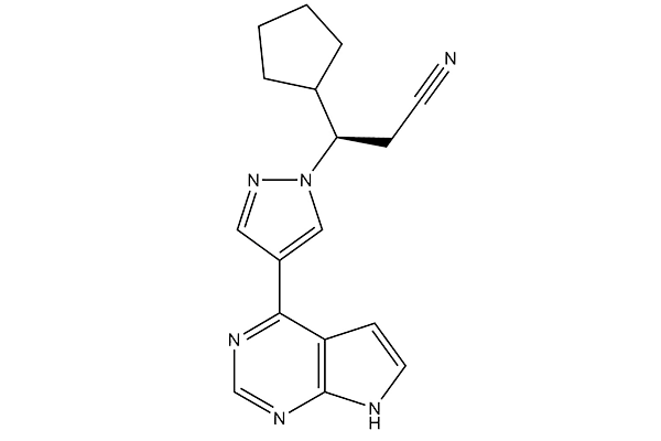
CAS No. 941678-49-5
文献中Selleckの製品使用例(641)
製品安全説明書
現在のバッチを見る:
純度:
99.98%
99.98
Ruxolitinib (INCB18424)と併用されることが多い化合物
It and Linifanib (ABT-869) combination have a synergistic effect on FMS-like tyrosine kinase 3 (FLT3) inhibition in acute myeloid leukemia (AML) patients.
It and TP-3654 combination exhibit a significantly greater reduction of bone marrow (BM) fibrosis in MPLW515L mice.
Ruxolitinib (INCB18424)関連製品
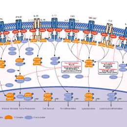
シグナル伝達経路
JAK阻害剤の選択性比較
Cell Data
| Cell Lines | Assay Type | Concentration | Incubation Time | 活性情報 | PMID |
|---|---|---|---|---|---|
| A549/DDP | Function Assay | 30 nM | 48 h | Down-regulation of STAT3 phosphorylation | 25213670 |
| NCI-H2347 | Function Assay | 30 nM | 48 h | Decrease in Bcl2 expression | 25213670 |
| NCI-H1299 | Function Assay | 30 nM | 48 h | Down-regulation of STAT3 phosphorylation | 25213670 |
| A549/DDP | Apoptosis Assay | 30 nM | 48 h | Induction of apoptosis | 25213670 |
| NCI-H1299 | Apoptosis Assay | 30 nM | 48 h | Induction of apoptosis | 25213670 |
| NCI-H2347 | Apoptosis Assay | 30 nM | 48 h | Induction of apoptosis | 25213670 |
| Hep3B | Function Assay | 1 μM | 16 h | Impaires the capacity of IHCA-associated gp130 mutants to active STAT3 with IC50 of ~50 μM | 24501689 |
| HepG2 | Function Assay | 1 μM | 16 h | Impaires the capacity of IHCA-associated gp130 mutants to signal to STAT3 | 24501689 |
| Huh7 | Function Assay | 1 μM | 16 h | Impaires the capacity of IHCA-associated gp130 mutants to signal to STAT3 | 24501689 |
| BaF3 | Kinase Assay | 80 nM | 6 h | Reduces the phosphorylation of STAT5 in JAK2V617F-mutated BAF3-EPOR cell | 24237791 |
| DLD-1 | Kinase Assay | 25 μM | 48 h | Inhibition of JAK1 phosphorylation | 24050550 |
| RKO | Kinase Assay | 25 μM | 48 h | Inhibition of JAK1 phosphorylation | 24050550 |
| DLD-1 | Kinase Assay | 25 μM | 48 h | Inhibition of JAK2 phosphorylation | 24050550 |
| RKO | Kinase Assay | 25 μM | 48 h | does not inhibit JAK1 phosphorylation | 24050550 |
| RKO | Growth Inhibition Assay | 50 μM | 48 h | IC50=14.76 μM | 24050550 |
| DLD-1 | Growth Inhibition Assay | 50 μM | 48 h | IC50=15.51 μM | 24050550 |
| DLD-1 | Apoptosis Assay | 25 μM | 48 h | Induces apoptosis by activating caspase 3 | 24050550 |
| RKO | Apoptosis Assay | 25 μM | 48 h | Induces apoptosis by activating caspase 3 | 24050550 |
| HuH7 | Growth Inhibition Assay | 50 μM | 48 h | >82% reduction | 23941832 |
| SNU182 | Growth Inhibition Assay | 50 μM | 48 h | >64% reduction | 23941832 |
| SNU423 | Growth Inhibition Assay | 50 μM | 48 h | >81% reduction | 23941832 |
| HuH7 | Function Assay | 50 μM | 24 h | Inhibition of STAT1 and STAT3 phosphorylation significantly | 23941832 |
| SNU182 | Function Assay | 50 μM | 24 h | Inhibition of STAT1 and STAT3 phosphorylation significantly | 23941832 |
| SNU423 | Function Assay | 50 μM | 24 h | Inhibition of STAT1 and STAT3 phosphorylation significantly | 23941832 |
| HT93A | Growth Inhibition Assay | 320 nM | 5 d | Inhibition of GCS-F induced granulocytic differentiation | 25805962 |
| SET-2 | Cytotoxic Assay | 5 μM | 48 h | Cytotoxic index=18.7% | 25931349 |
| HEL | Cytotoxic Assay | 5 μM | 48 h | Cytotoxic index=12.2% | 25931349 |
| TF1 | Kinase Assay | 20 min | Inhibition of JAK1 in human TF1 cells assessed as inhibition of IL6-induced STAT3 phosphorylation with IC50 of 0.024μM | 22698084 | |
| TF1 | Kinase Assay | 20 min | Inhibition of JAK2 in human TF1 cells assessed as inhibition of EPO-induced STAT5 phosphorylation with IC50 of 0.012μM | 22698084 | |
| Sf9 cells | JAK inhibition assay | 1 h | Ki = 0.0001 μM | 23668484 | |
| Sf9 cells | JAK inhibition assay | 1 h | Ki = 0.0002 μM | 23668484 | |
| Sf9 cells | JAK inhibition assay | 1 h | Ki = 0.0005 μM | 23668484 | |
| Sf21 cells | JAK inhibition assay | 1 h | IC50 = 0.0028 μM | 22591402 | |
| Sf21 cells | JAK inhibition assay | 60 min | IC50 = 0.003 μM | 27137359 | |
| Sf9 cells | JAK inhibition assay | 1 h | Ki = 0.0032 μM | 23668484 | |
| Sf21 cells | JAK inhibition assay | 1 h | IC50 = 0.0033 μM | 22591402 | |
| TF1 cells | JAK inhibition assay | 30 min | IC50 = 0.00685 μM | 23061660 | |
| TF1 cells | JAK inhibition assay | 20 min | EC50 = 0.012 μM | 22698084 | |
| Sf21 cells | TYK2 inhibition assay | 1 h | IC50 = 0.019 μM | 22591402 | |
| TF1 cells | JAK inhibition assay | 20 min | EC50 = 0.024 μM | 22698084 | |
| Sf21 cells | JAK inhibition assay | 1 h | IC50 = 0.428 μM | 22591402 | |
| CD34+ cells | JAK inhibition assay | 45 min | IC50 = 0.677 μM | 24417533 | |
| NCI-H2347 | Growth Inhibition Assay | IC50=0.17 μM | 25213670 | ||
| NCI-H1299 | Growth Inhibition Assay | IC50=0.28 μM | 25213670 | ||
| A549/DDP | Growth Inhibition Assay | IC50=0.22 μM | 25213670 | ||
| A549 | Growth Inhibition Assay | IC50=0.04 μM | 25213670 | ||
| NCI-H358 | Growth Inhibition Assay | IC50=0.1 μM | 25213670 | ||
| NCI-H460 | Growth Inhibition Assay | IC50=0.13 μM | 25213670 | ||
| CMK | Growth Inhibition Assay | Inhibition of CMK carrying the WT JAK cell proliferation with IC50 of 0.075 μM | 25352124 | ||
| CMK | Growth Inhibition Assay | Inhibition of CMK carrying the JAK3A63D mutation cell proliferation with IC50 of 0.163 μM | 25352124 | ||
| CMK | Growth Inhibition Assay | Inhibition of CMK carrying the JAK3A572V mutation cell proliferation | 25352124 | ||
| Human monocyte | Kinase Assay | Inhibition of JAK2/1 in human monocytes expressing CD14 assessed as inhibition of IFNgamma-stimulated STAT1 phosphorylation with IC50 of 0.031μM | 23540648 | ||
| Human monocyte | Kinase Assay | Inhibition of JAK2 in human monocytes expressing CD14 assessed as inhibition of GM-CSF-stimulated STAT5a phosphorylation with IC50 of 0.026μM | 23540648 | ||
| Human T cell | Kinase Assay | Inhibition of JAK3/1 in human T cells expressing CD3 assessed as inhibition of IL2-stimulated STAT5a phosphorylation with IC50 of 0.023μM | 23540648 | ||
| SET2 cells | JAK inhibition assay | IC50 = 0.00184 μM | 23061660 | ||
| CD34+ cells | JAK inhibition assay | IC50 = 0.008 μM | 26927423 | ||
| T cells | JAK inhibition assay | IC50 = 0.023 μM | 23540648 | ||
| T cells | JAK inhibition assay | IC50 = 0.023 μM | 23540648 | ||
| T cells | JAK inhibition assay | IC50 = 0.031 μM | 23540648 | ||
| T cells | JAK inhibition assay | IC50 = 0.031 μM | 23540648 | ||
| PBMC cells | JAK inhibition assay | IC50 = 0.04 μM | 26927423 | ||
| PBMC cells | STAT5 inhibition assay | IC50 = 0.448 μM | 26927423 | ||
| 他の多くの細胞株試験データをご覧になる場合はこちらをクリックして下さい | |||||
生物活性
| 製品説明 | ルクソリチニブ (Ruxolitinib (INCB018424)) は、最初に臨床試験が行われた強力で選択的なJAK1/2 阻害剤であり、IC50 はそれぞれ 3.3 nM/ 2.8 nM、JAK1/2 に対して JAK3 の 130 倍の選択性を示します。ルクソリチニブはマイトファジー (mitophagy) によって抗腫瘍作用を示します。また、オートファジー (autophagy) を誘導し、アポトーシス (apoptosis) を促進します。 | ||||
|---|---|---|---|---|---|
| Targets |
|
| In Vitro | ||||
| In vitro | Ruxolitinib (INCB18424) potently and selectively inhibits JAK2V617F-mediated signaling and proliferation in Ba/F3 cells and HEL cells. This compound markedly increases apoptosis in a dose dependent manner in Ba/F3 cells. It (64 nM) results in a doubling of cells with depolarized mitochondria in Ba/F3 cells. This chemical inhibits proliferating of erythroid progenitors from normal donors and polycythemia vera patients with IC50 of 407 nM and 223 nM, respectively. It demonstrates remarkable potency against erythroid colony formation with IC50 of 67nM. | |||
|---|---|---|---|---|
| Kinase Assay | Binding assay | |||
| Recombinant proteins are expressed using Sf21 cells and baculovirus vectors and purified with affinity chromatography. JAK kinase assays use a homogeneous time-resolved fluorescence assay with the peptide substrate (-EQEDEPEGDYFEWLE). Each enzyme reaction is carried out with Ruxolitinib (INCB18424) or control, JAK enzyme, 500 nM peptide, adenosine triphosphate (ATP; 1mM), and 2% dimethyl sulfoxide (DMSO) for 1 hour. The 50% inhibitory concentration (IC50) is calculated as the concentration of this compound required for inhibition of 50% of the fluorescent signal. | ||||
| 細胞実験 | 細胞株 | Ba/F3 and HEL cells | ||
| 濃度 | 3 μM | |||
| 反応時間 | 48 hours | |||
| 実験の流れ | Cells are seeded at 2 × 10³/well of white bottom 96-well plates and treated with Ruxolitinib (INCB018424) from DMSO stocks (0.2% final DMSO concentration), followed by incubation for 48 hours at 37 ℃ with 5% CO2. Viability is measured by cellular ATP determination using the Cell-Titer Glo luciferase reagent or viable cell counting. Values are transformed to percent inhibition relative to vehicle control, and IC50 curves are fitted according to nonlinear regression analysis of the data using PRISM GraphPad. | |||
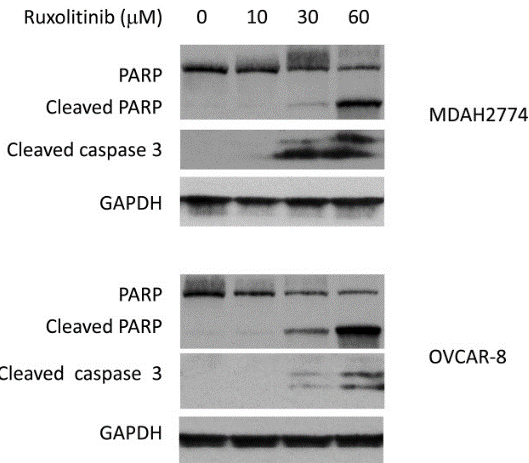
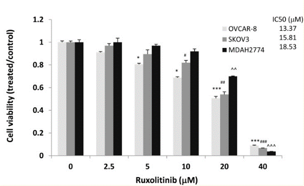
| 実験結果図 | Methods | Biomarkers | 結果図 | PMID |
| Western blot | cleaved PARP / cleaved caspase3 p-JAK2 / p-AKT / p-MAPK / Bcl-xl / MCL-1 c-Myc / c-Jun / Cyclin B / Cyclin D / Bcl-2 / HIF-1α p-STAT3 |
|
29849942 | |
| Growth inhibition assay | Cell viability Cell apoptosis Cell proliferation |
|
29849942 | |
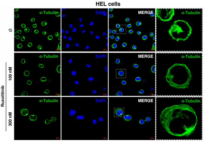
| Immunofluorescence | α-tubulin |
|
26356819 | |
| In Vivo | ||
| In Vivo | INCB018424 (180 mg/kg, orally, twice a day) results in a survival rate of greater than 90% by day 22 in a JAK2V617F-driven mouse model. This compound (180 mg/kg, orally, twice a day) markedly reduces splenomegaly and circulating levels of inflammatory cytokines, and preferentially eliminates neoplastic cells, resulting in significantly prolonged survival without myelosuppressive or immunosuppressive effects in a JAK2V617F-driven mouse model. In the double-blind trial of myelofibrosis, the primary end point is reached in 41.9% of patients in the Ruxolitinib (INCB18424) group as compared with 0.7% in the placebo group. It results in maintaining reduction in spleen volume and improvement of 50% or more in the total symptom score. A total of 28% of the patients in the group receiving this compound (15 mg twice daily) has at least a 35% reduction in spleen volume at week 48 in patients with myelofibrosis, as compared with 0% in the group receiving the best available therapy. The mean palpable spleen length has decreased by 56% with it but has increased by 4% with the best available therapy at week 48. Patients in the ruxolitinib group has an improvement in overall quality-of-life measures and a reduction in symptoms associated with myelofibrosis. | |
|---|---|---|
| 動物実験 | 動物モデル | JAK2V617F-driven mouse model |
| 投与量 | 180 mg/kg | |
| 投与経路 | Oral gavage | |
| NCT Number | Recruitment | Conditions | Sponsor/Collaborators | Start Date | Phases |
|---|---|---|---|---|---|
| NCT06397313 | Not yet recruiting | Myelofibrosis |
Ryvu Therapeutics SA |
September 2024 | Phase 2 |
| NCT06388564 | Not yet recruiting | Chronic Graft-versus-host-disease |
Incyte Corporation |
July 8 2024 | Phase 2 |
| NCT06251102 | Not yet recruiting | Polycythemia Vera |
Gruppo Italiano Malattie EMatologiche dell''Adulto |
July 2024 | -- |
| NCT06343792 | Not yet recruiting | Steroid Refractory GVHD |
ReAlta Life Sciences Inc. |
May 2024 | Phase 2 |
|
化学情報
| 分子量 | 306.37 | 化学式 | C17H18N6 |
| CAS No. | 941678-49-5 | SDF | Download Ruxolitinib (INCB18424) SDFをダウンロードする |
| Smiles | C1CCC(C1)C(CC#N)N2C=C(C=N2)C3=C4C=CNC4=NC=N3 | ||
| 保管 | 3 years-20°C(in the dark) powder | ||
|
In vitro |
DMSO : 300 mg/mL ( (979.2 mM); 吸湿したDMSOは溶解度を減少させます。新しいDMSOをご使用ください。) Ethanol : 12 mg/mL Water : Insoluble |
モル濃度計算器 |
|
in vivo Add solvents to the product individually and in order. |
投与溶液組成計算機 | |||||
実験計算
投与溶液組成計算機(クリア溶液)
ステップ1:実験データを入力してください。(実験操作によるロスを考慮し、動物数を1匹分多くして計算・調製することを推奨します)
mg/kg
g
μL
匹
ステップ2:投与溶媒の組成を入力してください。(ロット毎に適した溶解組成が異なる場合があります。詳細については弊社までお問い合わせください)
% DMSO
%
% Tween 80
% ddH2O
%DMSO
%
計算結果:
投与溶媒濃度: mg/ml;
DMSOストック溶液調製方法: mg 試薬を μL DMSOに溶解する(濃度 mg/mL, 注:濃度が当該ロットのDMSO溶解度を超える場合はご連絡ください。 )
投与溶媒調製方法:Take μL DMSOストック溶液に μL PEG300,を加え、完全溶解後μL Tween 80,を加えて完全溶解させた後 μL ddH2O,を加え完全に溶解させます。
投与溶媒調製方法:μL DMSOストック溶液に μL Corn oil,を加え、完全溶解。
注意:1.ストック溶液に沈殿、混濁などがないことをご確認ください;
2.順番通りに溶剤を加えてください。次のステップに進む前に溶液に沈殿、混濁などがないことを確認してから加えてください。ボルテックス、ソニケーション、水浴加熱など物理的な方法で溶解を早めることは可能です。
技術サポート
ストックの作り方、阻害剤の保管方法、細胞実験や動物実験の際に注意すべき点など、製品を取扱う時に問い合わせが多かった質問に対しては取扱説明書でお答えしています。
他に質問がある場合は、お気軽にお問い合わせください。
* 必須
よくある質問(FAQ)
質問1:
What is the difference between S2902 and S1378 which seem to have same structure formula according to the product information?
回答
These two chemicals are the two different chiral forms of this compound. S2902 S-Ruxolitinib is the S form and S1378 Ruxolitinib is the D form. One of the carbon atoms in it is asymmetric, making the two molecules mirror images of each other. The biological activities of these two molecules can be very different because of the confirmation differences.
質問2:
How about the half-life of this compound? How long is the duration of its inhibitory effect on JAK-STAT signaling?
回答
According to previous study, the half-life of this compound in body is about 2~3 hours. Generally, it is longer in vitro culture medium than in vivo. It was also used for 24 hours in paper. http://www.bloodjournal.org/cgi/pmidlookup?view=long&pmid=24711661.
納期 国内在庫品:受注日の翌日(15時までの受注分) *北海道、九州、沖縄への配送は受注日より2日以上 を要する場合あり 海外在庫品:受注後1〜2週間