
- 阻害剤
- 研究分野別
- PI3K/Akt/mTOR
- Epigenetics
- Methylation
- Immunology & Inflammation
- Protein Tyrosine Kinase
- Angiogenesis
- Apoptosis
- Autophagy
- ER stress & UPR
- JAK/STAT
- MAPK
- Cytoskeletal Signaling
- Cell Cycle
- TGF-beta/Smad
- 化合物ライブラリー
- 抗体
- 新製品
- お問い合わせ
Canertinib (CI-1033)
別名:PD183805
Canertinib (CI-1033, PD183805) は、IC50が1.5 nMと9.0 nMのEGFRおよびErbB2に対するパン-ErbB阻害剤であり、PDGFR、FGFR、InsR、PKC、またはCDK1/2/4には活性がありません。第3相。
CAS No. 267243-28-7
文献中Selleckの製品使用例(51)
製品安全説明書
現在のバッチを見る:
純度:
99.99%
99.99
Canertinib (CI-1033)関連製品
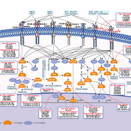
シグナル伝達経路
EGFR阻害剤の選択性比較
Cell Data
| Cell Lines | Assay Type | Concentration | Incubation Time | 活性情報 | PMID |
|---|---|---|---|---|---|
| human A431 cells | Function assay | 1 μM | 1 h | Irreversible inhibition of EGFR autophosphorylation in human A431 cells at 1 uM incubated for 1 hr followed by compound wash out measured 5 hrs post EGF addition by Western blotting analysis | |
| human LNCaP cells | Function assay | 10 μM | 2 h | Inhibition of autophosphorylation of immunoprecipitated flag-tagged Bmx expressed in human LNCaP cells assessed as incorporation of [32P]ATP at 10 uM pretreated for 2 hrs before transfection by immunoblot analysis | |
| human HCC827 cells | Proliferation assay | 72 h | Antiproliferative activity against human HCC827 cells harboring EGFR del E746-A750 mutant after 72 hrs by MTS assay, IC50=0.001 μM | ||
| human BT474 cells | Proliferation assay | 3 days | Antiproliferative activity against human BT474 cells overexpressing ERBb2 after 3 days by methylene blue staining, EC50=0.01 μM | ||
| human HN5 cells | Proliferation assay | 3 days | Antiproliferative activity against human HN5 cells overexpressing EGFR after 3 days by methylene blue staining, EC50=0.05 μM | ||
| human NCI-H1975 cells | Proliferation assay | 72 h | Antiproliferative activity against human NCI-H1975 cells harboring EGFR L858R/T790M mutant after 72 hrs by MTS assay, IC50=0.064 μM | ||
| human A431 cells | Proliferation assay | 72 h | Antiproliferative activity against human A431 cells overexpressing EGFR after 72 hrs by MTS assay, IC50=0.15 μM | ||
| human A549 cells | Proliferation assay | 72 h | Antiproliferative activity against human A549 cells expressing wild type EGFR coexpressing k-Ras mutant after 72 hrs by MTS assay, IC50=1.59 μM | ||
| human HL7702 cells | Proliferation assay | 72 h | Antiproliferative activity against human HL7702 cells expressing wilt type EGFR after 72 hrs by MTS assay, IC50=2.3 μM | ||
| NCI-H1975 cells | Growth inhibition assay | 48 h | Inhibition of EGFR L858R/T790M mutant in human NCI-H1975 cells assessed as growth inhibition after 48 hrs by MTT assay | ||
| A431 cells | Function assay | Inhibition of EGF-stimulated autophosphorylation of EGFR enzyme in A431 cells detected by immunoblotting, IC50=0.0074 μM | |||
| MDA-MB 453 cells | Function assay | Inhibition of autophosphorylation of ERBB2 receptor kinase in MDA-MB 453 cells, IC50=0.009 μM | |||
| mouse BAF3 cells | Function assay | Inhibition of Blk expressed in mouse BAF3 cells assessed as cytotoxicity, IC50=0.029 μM | |||
| mouse BAF3 cells | Function assay | Inhibition of JAK3 expressed in mouse BAF3 cells assessed as cytotoxicity, IC50=2 μM | |||
| 他の多くの細胞株試験データをご覧になる場合はこちらをクリックして下さい | |||||
生物活性
| 製品説明 | Canertinib (CI-1033, PD183805) は、IC50が1.5 nMと9.0 nMのEGFRおよびErbB2に対するパン-ErbB阻害剤であり、PDGFR、FGFR、InsR、PKC、またはCDK1/2/4には活性がありません。第3相。 | ||||
|---|---|---|---|---|---|
| 特性 | First kinase inhibitor to show irreversible activity and to have entered clinical trials (serving as a template for further development). | ||||
| Targets |
|
| In Vitro | ||||
| In vitro |
Canertinib (CI-1033) shows excellent potency for irreversible inhibition of erbB2 autophosphorylation in MDA-MB 453 cells, and also demonstrates high permeability in Caco-2 cells. This compound alone significantly suppresses constitutively activated Akt and MAP kinase, and in combination inhibits Akt while preventing increased levels of MAPK phosphorylation. It stimulates p27 expression and p38 phosphorylation in MDA-MB-453 cells. CI-1033 is highly specific to the erbB receptor family and not sensitive to PGFR, FGFR or IR even at 50 μM. It shows high levels of inhibition in A431 cells expressing EGFR with IC50 of 7.4 nM, and suppresses heregulin-stimulated tyrosine phosphorylation of erbB2, erbB3 and erbB4 with IC50 of 5, 14 and 10 nM, respectively. The compound also inhibits expression of pp62c-fos in response to heregulin. It is predicted to modify Cys773 covalently within the ATP binding site of the HER2 kinase and enhances destruction of both mature and immature ErbB-2 molecules. This compound induces a significant decrease in measurable phosphorylation of tyrosine residues 845 and 1068 of EGFR, which are responsible for Src and Ras/MAPK signaling respectively. The corresponding residues of Her-2, tyrosine residues 877 and 1248 are dephosphorylated significantly by it at a concentration of 3 μM or higher. CI could block EGFR internalization and increase the rate of apoptosis in primary osteosarcoma cells in a titratable fashion. In addition, it inhibits the proliferation of TT, TE2, TE6 and TE10 cells significantly at 0.1 nM. |
|||
|---|---|---|---|---|
| Kinase Assay | Tyrosine Kinase Assays | |||
| Enzyme assays for determination of IC50 are performed in 96-well filter plates in a total volume of 0.1 mL, containing 20 mM Hepes, pH 7.4, 50 mM sodium vanadate, 10 μM the ATP containing 0.5 mCi of [32P]ATP, 20 mg of polyglutamic acid/tyrosine, 10 ng of EGFR tyrosine kinase, and appropriate dilutions of Canertinib (CI-1033). All components except the ATP are added to the well and the plate is incubated with shaking for 10 min at 25 °C. The reaction is started by adding [32P]ATP, and the plate is incubated at 25 °C for another 10 min. The reaction is terminated by addition of 0.1 mL of 20% trichloroacetic acid (TCA). The plate is kept at 4 °C for at least 15 min to allow the substrate to precipitate. The wells are then washed five times with 0.2 mL of 10% TCA and 32P incorporation determined with a Wallac β plate counter. | ||||
| 細胞実験 | 細胞株 | TT, TE2, TE6 and TE10 cells | ||
| 濃度 | 0.1-5.0 nM | |||
| 反応時間 | 1, 3, 5 and 7 days | |||
| 実験の流れ | Cells (1 × 10⁴) are seeded in each well of a 24-well plastic culture plate and left overnight in DMEM or RPMI-1640 supplemented with 10% FBS. The next morning, the cells are treated with the indicated concentrations of Canertinib (CI-1033) (0.1-5.0 nM) for varying periods (1, 3, 5 and 7 days). After treatment, the cells are counted using a Coulter counter. The percent of cell proliferation is calculated by this formula: treatment cell number/control cell number × 100 for each time period. |
|||
| 実験結果図 | Methods | Biomarkers | 結果図 | PMID |
| Western blot | pEGFR / EGFR / p-HER2 / HER2 / p-HER3 / HER3 / MUC4 p-FAK / FAK / p-AKT / AKT |
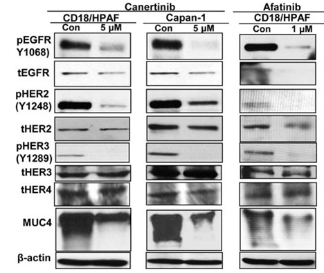
|
25686822 | |
| Growth inhibition assay | Cell viability |
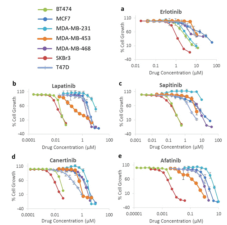
|
28638122 | |
| In Vivo | ||
| In Vivo |
Canertinib (CI-1033) shows impressive activity against A431 xenografts in nude mice at 5 mg/kg of body weight. This compound (20 to 80 mg/kg/d) achieves a high degree of tumor regressions in H125 xenograft models. Its oral administration causes a marked inhibition of growth in TT, TE6 and TE10 xenografts in nude mice, without animal death and <10% weight loss. |
|
|---|---|---|
| 動物実験 | 動物モデル | A431 xenografts established in nude mice |
| 投与量 | ~18 mg/kg | |
| 投与経路 | Administered orally | |
| NCT Number | Recruitment | Conditions | Sponsor/Collaborators | Start Date | Phases |
|---|---|---|---|---|---|
| NCT00050830 | Completed | Lung Neoplasms |
Pfizer |
January 2003 | Phase 2 |
| NCT00174356 | Completed | Carcinoma Non-Small Cell Lung |
Pfizer |
December 2002 | Phase 1 |
| NCT00051051 | Completed | Breast Neoplasms |
Pfizer |
December 2002 | Phase 2 |
|
化学情報
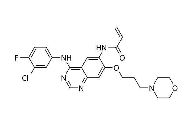
| 分子量 | 485.94 | 化学式 | C24H25ClFN5O3 |
| CAS No. | 267243-28-7 | SDF | Download Canertinib (CI-1033) SDFをダウンロードする |
| Smiles | C=CC(=O)NC1=C(C=C2C(=C1)C(=NC=N2)NC3=CC(=C(C=C3)F)Cl)OCCCN4CCOCC4 | ||
| 保管 | |||
|
In vitro |
4-Methylpyridine : 100 mg/mL DMSO : Insoluble ( 吸湿したDMSOは溶解度を減少させます。新しいDMSOをご使用ください。) Water : Insoluble |
モル濃度計算器 |
|
in vivo Add solvents to the product individually and in order. |
投与溶液組成計算機 | |||||
実験計算
投与溶液組成計算機(クリア溶液)
ステップ1:実験データを入力してください。(実験操作によるロスを考慮し、動物数を1匹分多くして計算・調製することを推奨します)
mg/kg
g
μL
匹
ステップ2:投与溶媒の組成を入力してください。(ロット毎に適した溶解組成が異なる場合があります。詳細については弊社までお問い合わせください)
% DMSO
%
% Tween 80
% ddH2O
%DMSO
%
計算結果:
投与溶媒濃度: mg/ml;
DMSOストック溶液調製方法: mg 試薬を μL DMSOに溶解する(濃度 mg/mL, 注:濃度が当該ロットのDMSO溶解度を超える場合はご連絡ください。 )
投与溶媒調製方法:Take μL DMSOストック溶液に μL PEG300,を加え、完全溶解後μL Tween 80,を加えて完全溶解させた後 μL ddH2O,を加え完全に溶解させます。
投与溶媒調製方法:μL DMSOストック溶液に μL Corn oil,を加え、完全溶解。
注意:1.ストック溶液に沈殿、混濁などがないことをご確認ください;
2.順番通りに溶剤を加えてください。次のステップに進む前に溶液に沈殿、混濁などがないことを確認してから加えてください。ボルテックス、ソニケーション、水浴加熱など物理的な方法で溶解を早めることは可能です。
技術サポート
ストックの作り方、阻害剤の保管方法、細胞実験や動物実験の際に注意すべき点など、製品を取扱う時に問い合わせが多かった質問に対しては取扱説明書でお答えしています。
他に質問がある場合は、お気軽にお問い合わせください。
* 必須
よくある質問(FAQ)
質問1:
I would like to know which is the best option/solvent to dilute it for in vivo experiments. (I am treating mice at 30mg/mL of this compound.)
回答
It is a suspension in the formulation recommended (30% Propylene glycol, 5% Tween 80, 65% D5W) on our product page at 30mg/ml. It’s fine for oral gavage.
納期 国内在庫品:受注日の翌日(15時までの受注分) *北海道、九州、沖縄への配送は受注日より2日以上 を要する場合あり 海外在庫品:受注後1〜2週間