
- 阻害剤
- 研究分野別
- PI3K/Akt/mTOR
- Epigenetics
- Methylation
- Immunology & Inflammation
- Protein Tyrosine Kinase
- Angiogenesis
- Apoptosis
- Autophagy
- ER stress & UPR
- JAK/STAT
- MAPK
- Cytoskeletal Signaling
- Cell Cycle
- TGF-beta/Smad
- 化合物ライブラリー
- 抗体
- 新製品
- お問い合わせ
Tubastatin A
Tubastatin Aは、無細胞アッセイで15nMのIC50を持つ強力で選択的なHDAC6阻害剤です。HDAC8(57倍)を除く他のすべてのアイソザイムに対して選択性があります(1000倍)。Tubastatin Aはオートファジーを促進し、アポトーシスを増加させます。
CAS No. 1252003-15-8
文献中Selleckの製品使用例(111)
製品安全説明書
現在のバッチを見る:
純度:
99.65%
99.65
Tubastatin A関連製品
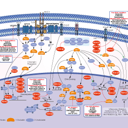
シグナル伝達経路
HDAC阻害剤の選択性比較
Cell Data
| Cell Lines | Assay Type | Concentration | Incubation Time | 活性情報 | PMID |
|---|---|---|---|---|---|
| KB | Function assay | 14 uM | 24 hrs | Inhibition of HDAC1 in human KB cells assessed as increase in histone H4 acetylation at 14 uM after 24 hrs by Western blotting analysis | 25899338 |
| KMS-12-BM | Function assay | 15 uM | up to 48 hrs | Inhibition of HDAC6 in human KMS-12-BM cells assessed as increase in acetylated tubulin level at 15 uM up to 48 hrs by immunoblot method | 27541357 |
| MOLM14 | Function assay | 15 uM | up to 48 hrs | Inhibition of HDAC6 in human MOLM14 cells assessed as increase in acetylated tubulin level at 15 uM up to 48 hrs by immunoblot method | 27541357 |
| U937 | Function assay | 2 uM | 18 hrs | Inhibition of HDAC6 in human U937 cells assessed as increase in alpha-tubulin acetylation at Lys-40 residue at 2 uM after 18 hrs by Western blot method | 28337317 |
| HEL | Cell cycle assay | 1 to 5 uM | 48 hrs | Cell cycle arrest in human HEL cells assessed as accumulation at G1 phase at 1 to 5 uM after 48 hrs propidium iodide staining based flow cytometry | 29940115 |
| HeLa | Function assay | 2 uM | 12 hrs | Inhibition of HDAC6 in human HeLa cells assessed as increase in acetyl-tubulin level at 2 uM after 12 hrs by Western blot analysis | 29533873 |
| HEK293 | Function assay | 10 uM | 24 hrs | Inhibition of HDAC1 in HEK293 cells assessed as increase in histone H3 acetylation at 10 uM after 24 hrs by Western blot method | 28523102 |
| MV4-11 | Function assay | 200 nM | 24 hrs | Inhibition of HDAC6 in human MV4-11 cells assessed as accumulation of acetylated alpha-tubulin at 200 nM after 24 hrs by Western blot analysis | 29738953 |
| HCT116 | Cell cycle assay | 5 uM | 48 hrs | Cell cycle arrest in human HCT116 cells assessed as accumulation at sub-G1 phase at 5 uM after 48 hrs by propidium iodide staining-based flow cytometric method | 28038324 |
| PC12 | Neuroprotective assay | 10 uM | 24 hrs | Neuroprotective activity against H2O2-induced toxicity in rat PC12 cells assessed as cell viability at 10 uM pretreated for 24 hrs followed by H2O2 challenge and measured after 12 hrs by MTT assay relative to control | 30385227 |
| PC12 | Neuroprotective assay | 5 uM | 24 hrs | Neuroprotective activity against 6-OHDA-induced toxicity in rat PC12 cells assessed as increase in cell viability at 5 uM pretreated for 24 hrs followed by 6-OHDA challenge and measured after 12 hrs by MTT assay | 30385227 |
| PC12 | Neuroprotective assay | 10 uM | 24 hrs | Neuroprotective activity against 6-OHDA-induced toxicity in rat PC12 cells assessed as increase in cell viability at 10 uM pretreated for 24 hrs followed by 6-OHDA challenge and measured after 12 hrs by MTT assay | 30385227 |
| PC12 | Neuroprotective assay | 5 to 10 uM | 24 hrs | Neuroprotective activity against 6-OHDA-induced toxicity in rat PC12 cells assessed as increase in cell viability at 5 to 10 uM pretreated for 24 hrs followed by 6-OHDA challenge and measured after 12 hrs coincubated with ebselen by MTT assay | 30385227 |
| PC12 | Antioxidant assay | 5 uM | 24 hrs | Antioxidant activity against H2O2-induced oxidative stress in rat PC12 cells assessed as decrease in ROS accumulation at 5 uM preincubated for 24 hrs followed by H2O2 challenge and measured after 12 hrs by DCFH-DA dye-based fluorescence analysis | 30385227 |
| PC12 | Antioxidant assay | 5 uM | 24 hrs | Antioxidant activity against H2O2-induced oxidative stress in rat PC12 cells assessed as decrease in ROS accumulation at 5 uM preincubated for 24 hrs followed by H2O2 challenge and measured after 12 hrs coincubated with ebselen by DCFH-DA dye-based fluore | 30385227 |
| Sf9 | Function assay | 30 mins | Inhibition of full length human recombinant N-terminal GST-tagged HDAC6 expressed in baculovirus infected sf9 cells using fluorogenic HDAC substrate 3 after 30 mins by fluorescence assay, IC50 = 0.013 μM. | 29549837 | |
| Sf9 | Function assay | 2 hrs | Inhibition of full length human recombinant N-terminal GST-tagged HDAC6 (1 to 1215 residues) expressed in sf9 cells preincubated with enzyme followed by fluorogenic Arg-His-Lys-Lys(Ac)-AMC substrate addition measured after 2 hrs by fluorescence assay, IC50 = 0.0035 μM. | 27541357 | |
| Sf9 | Function assay | 90 mins | Inhibition of full length human recombinant N-terminal GST-tagged HDAC6 expressed in Sf9 cells using RHKK(Ac) as substrate after 90 mins by fluorimetric method, IC50 = 0.0137 μM. | 28038324 | |
| Sf9 | Function assay | 2 hrs | Inhibition of human recombinant HDAC6 expressed in Sf9 cells incubated for 2 hrs using RHKK-Ac fluorogenic substrate, IC50 = 0.015 μM. | 23009203 | |
| Sf9 | Function assay | 90 mins | Inhibition of human recombinant N-terminal GST-tagged full length HDAC6 expressed in insect SF9 cells using fluorogenic ZMAL as substrate after 90 mins by fluorescence-based assay, IC50 = 0.0304 μM. | 30092367 | |
| HeLaS3 | Function assay | 15 mins | Inhibition of HDAC6 in human HeLaS3 cells preincubated for 15 mins followed by HDAC-Glo substrate addition measured after 30 to 45 mins by ELISA, IC50 = 0.031 μM. | 28337317 | |
| insect cells | Function assay | 4 hrs | Inhibition of human recombinant HDAC6 expressed in baculovirus infected insect cells using BATCP as substrate after 4 hrs by UHPLC-ESI-MS/MS analysis, IC50 = 0.0349 μM. | 27650925 | |
| SHSY5Y | Function assay | 8 hrs | Inhibition of HDAC6 in human SHSY5Y cells using BATCP as substrate after 8 hrs by UHPLC-ESI-MS/MS analysis, IC50 = 0.0943 μM. | 27650925 | |
| SHSY5Y | Function assay | 8 hrs | Inhibition of HDAC in human SHSY5Y cells using MAL as substrate after 8 hrs by UHPLC-ESI-MS/MS analysis, IC50 = 0.1221 μM. | 27650925 | |
| Sf9 | Function assay | 4 hrs | Inhibition of full length human recombinant C-terminal FLAG/His-tagged HDAC1 expressed in baculovirus infected sf9 cells using fluorogenic HDAC substrate 3 after 4 hrs fluorescence assay, IC50 = 0.718 μM. | 29549837 | |
| Sf9 | Function assay | 1 hr | Inhibition of full length human recombinant C-terminal FLAG/His-tagged HDAC1 expressed in baculovirus infected sf9 cells using fluorogenic HDAC substrate 3 after 1 hr fluorescence assay, IC50 = 0.967 μM. | 29549837 | |
| SHSY5Y | Function assay | 8 hrs | Inhibition of HDAC1 in human SHSY5Y cells using MOCPAC as substrate after 8 hrs by UHPLC-ESI-MS/MS analysis, IC50 = 1.1097 μM. | 27650925 | |
| Sf9 | Function assay | 30 mins | Inhibition of full length human recombinant C-terminal FLAG/His-tagged HDAC1 expressed in baculovirus infected sf9 cells using fluorogenic HDAC substrate 3 after 30 mins by fluorescence assay, IC50 = 1.54 μM. | 29549837 | |
| Sf9 | Function assay | 90 mins | Inhibition of human recombinant C-terminal His/FLAG-tagged full length HDAC1 expressed in insect SF9 cells using fluorogenic ZMAL as substrate after 90 mins by fluorescence-based assay, IC50 = 1.91 μM. | 30092367 | |
| HCT116 | Antiproliferative assay | 72 hrs | Antiproliferative activity against human HCT116 cells after 72 hrs by MTT assay, IC50 = 2 μM. | 27541357 | |
| Sf9 | Function assay | 30 mins | Inhibition of full length human recombinant C-terminal His-tagged HDAC3/N-terminal GST-tagged NCOR2 (95 to 489 residues) expressed in baculovirus infected sf9 cells using fluorogenic HDAC substrate 3 after 30 mins by fluorescence assay, IC50 = 2.26 μM. | 29549837 | |
| HeLa | Function assay | 6 hrs | Inhibition of HDAC6 in human HeLa cells assessed as reduction in K40 hyperacetylation of alpha-tubulin incubated for 6 hrs by immunofluorescence assay, IC50 = 2.5 μM. | 25454270 | |
| HL60 | Antiproliferative assay | 48 hrs | Antiproliferative activity against human HL60 cells after 48 hrs in presence of JAK2 inhibitor CYT-387 by CCK-8 assay, IC50 = 2.54 μM. | 29940115 | |
| K562 | Antiproliferative assay | 48 hrs | Antiproliferative activity against human K562 cells after 48 hrs in presence of JAK2 inhibitor CYT-387 by CCK-8 assay, IC50 = 2.54 μM. | 29940115 | |
| HEL | Antiproliferative assay | 48 hrs | Antiproliferative activity against human HEL cells after 48 hrs in presence of JAK2 inhibitor CYT-387 by CCK-8 assay, IC50 = 2.54 μM. | 29940115 | |
| HeLaS3 | Function assay | 15 mins | Inhibition of HDAC1 in human HeLaS3 cells preincubated for 15 mins followed by HDAC-Glo substrate addition measured after 30 to 45 mins by ELISA, IC50 = 2.7 μM. | 28337317 | |
| HeLaS3 | Function assay | 15 mins | Inhibition of HDAC3 in human HeLaS3 cells preincubated for 15 mins followed by HDAC-Glo substrate addition measured after 30 to 45 mins by ELISA, IC50 = 2.9 μM. | 28337317 | |
| Jurkat | Cytotoxicity assay | 72 hrs | Cytotoxicity against human Jurkat cells assessed as growth inhibition after 72 hrs by MTS assay, IC50 = 3.38 μM. | 24304348 | |
| MCF7 | Antiproliferative assay | 72 hrs | Antiproliferative activity against human MCF7 cells after 72 hrs by MTT assay, IC50 = 3.7 μM. | 27541357 | |
| HL60 | Antiproliferative assay | 48 hrs | Antiproliferative activity against human HL60 cells after 48 hrs by CCK-8 assay, IC50 = 3.75 μM. | 29940115 | |
| K562 | Antiproliferative assay | 48 hrs | Antiproliferative activity against human K562 cells after 48 hrs by CCK-8 assay, IC50 = 3.75 μM. | 29940115 | |
| HEL | Antiproliferative assay | 48 hrs | Antiproliferative activity against human HEL cells after 48 hrs by CCK-8 assay, IC50 = 3.75 μM. | 29940115 | |
| HeLaS3 | Function assay | 15 mins | Inhibition of HDAC2 in human HeLaS3 cells preincubated for 15 mins followed by HDAC-Glo substrate addition measured after 30 to 45 mins by ELISA, IC50 = 3.9 μM. | 28337317 | |
| CAL27 | Antiproliferative assay | 72 hrs | Antiproliferative activity against human CAL27 cells measured after 72 hrs by MTT assay, IC50 = 4.6 μM. | 28581289 | |
| PC3 | Antiproliferative assay | 72 hrs | Antiproliferative activity against human PC3 cells after 72 hrs by MTT assay, IC50 = 8.6 μM. | 27541357 | |
| Sf9 | Function assay | 30 mins | Inhibition of full length human recombinant C-terminal His-tagged HDAC2 expressed in baculovirus infected sf9 cells using fluorogenic HDAC substrate 3 after 30 mins by fluorescence assay, IC50 = 9.97 μM. | 29549837 | |
| MDA-MB-231 | Antiproliferative assay | 72 hrs | Antiproliferative activity against human MDA-MB-231 cells after 72 hrs by MTT assay, IC50 = 10.4 μM. | 27541357 | |
| Cal27CisR | Antiproliferative assay | 72 hrs | Antiproliferative activity against human Cal27CisR cells measured after 72 hrs by MTT assay, IC50 = 10.8 μM. | 28581289 | |
| LNCAP | Cytotoxicity assay | 72 hrs | Cytotoxicity against androgen-dependent human LNCAP cells assessed as growth inhibition after 72 hrs by MTS assay, IC50 = 10.88 μM. | 24304348 | |
| Cal27CisR | Function assay | 18 hrs | Inhibition of HDAC in human Cal27CisR cells using Boc-Lys(epsilon-Ac)-AMC as substrate preincubated for 18 hrs followed by substrate addition measured after 3 hrs by fluorescence assay, IC50 = 12.1 μM. | 28581289 | |
| KB | Cytotoxicity assay | 72 hrs | Cytotoxicity against human KB cells after 72 hrs by MTS assay, IC50 = 14.81 μM. | 25899338 | |
| THLE2 | Cytotoxicity assay | 72 hrs | Cytotoxicity against human THLE2 cells after 72 hrs by vialight cell proliferation assay, LC50 = 15.1 μM. | 29549837 | |
| CAL27 | Function assay | 18 hrs | Inhibition of HDAC in human CAL27 cells using Boc-Lys(epsilon-Ac)-AMC as substrate preincubated for 18 hrs followed by substrate addition measured after 3 hrs by fluorescence assay, IC50 = 16.1 μM. | 28581289 | |
| Sf9 | Function assay | 2 hrs | Inhibition of human recombinant HDAC1 expressed in Sf9 cells incubated for 2 hrs using RHKK-Ac fluorogenic substrate, IC50 = 16.4 μM. | 23009203 | |
| B16 | Growth inhibition assay | 48 hrs | Growth inhibition of mouse B16 cells incubated for 48 hrs by MTT assay, GI50 = 40.5 μM. | 23009203 | |
| LNCAP | Function assay | 24 hrs | Inhibition of HDAC6 in human LNCAP cells assessed as inhibition of DHT-induced alpha-tubulin deacetylation by measuring increase in alpha-tubulin acetylation measured after 24 hrs relative to control | 27717544 | |
| human | Function assay | 24 hrs | Antagonist activity at AR in human LNCAP cells assessed as suppression of DHT-induced AR protein level measured after 24 hrs relative to control | 27717544 | |
| PC12 | Cytoprotective assay | 24 hrs | Cytoprotective activity against H2O2-induced damage in rat PC12 cells assessed as decrease in ROS accumulation preincubated for 24 hrs followed by H2O2 challenge measured after 12 hrs by DCFH-DA dye-based fluorescence analysis | 30385227 | |
| PC12 | Cytoprotective assay | 24 hrs | Cytoprotective activity against H2O2-induced damage in rat PC12 cells assessed as decrease in ROS accumulation preincubated for 24 hrs followed by H2O2 challenge measured after 12 hrs by DCFH-DA dye-based inverted fluorescence microscopic analysis | 30385227 | |
| Sf9 | Function assay | Inhibition of full length human recombinant N-terminal GST-tagged HDAC6 (1 to 1215 residues) expressed in sf9 cells using RHK-K(Ac)-AMC as substrate by fluorescence assay, IC50 = 0.011 μM. | 27541357 | ||
| Sf9 | Function assay | Inhibition of human recombinant HDAC6 expressed in baculovirus/sf9 cells using RHKKAc as substrate, IC50 = 0.015 μM. | 23905680 | ||
| Sf9 | Function assay | Inhibition of human recombinant HDAC8 expressed in baculovirus/sf9 cells using RHKAcKAc as substrate, IC50 = 0.854 μM. | 23905680 | ||
| HCT116 | Antiproliferative assay | Antiproliferative activity against human HCT116 cells, IC50 = 2 μM. | 29945795 | ||
| MCF7 | Antiproliferative assay | Antiproliferative activity against human MCF7 cells, IC50 = 3.7 μM. | 29945795 | ||
| PC3 | Antiproliferative assay | Antiproliferative activity against human PC3 cells, IC50 = 8.6 μM. | 29945795 | ||
| MDA-MB-231 | Antiproliferative assay | Antiproliferative activity against human MDA-MB-231 cells, IC50 = 10.4 μM. | 29945795 | ||
| Sf9 | Function assay | Inhibition of human recombinant HDAC1 expressed in baculovirus/sf9 cells using RHKKAc as substrate, IC50 = 16.4 μM. | 23905680 | ||
| DAOY | qHTS assay | qHTS of pediatric cancer cell lines to identify multiple opportunities for drug repurposing: Primary screen for DAOY cells | 29435139 | ||
| SJ-GBM2 | qHTS assay | qHTS of pediatric cancer cell lines to identify multiple opportunities for drug repurposing: Primary screen for SJ-GBM2 cells | 29435139 | ||
| A673 | qHTS assay | qHTS of pediatric cancer cell lines to identify multiple opportunities for drug repurposing: Primary screen for A673 cells | 29435139 | ||
| SK-N-MC | qHTS assay | qHTS of pediatric cancer cell lines to identify multiple opportunities for drug repurposing: Primary screen for SK-N-MC cells | 29435139 | ||
| BT-37 | qHTS assay | qHTS of pediatric cancer cell lines to identify multiple opportunities for drug repurposing: Primary screen for BT-37 cells | 29435139 | ||
| NB-EBc1 | qHTS assay | qHTS of pediatric cancer cell lines to identify multiple opportunities for drug repurposing: Primary screen for NB-EBc1 cells | 29435139 | ||
| U-2 OS | qHTS assay | qHTS of pediatric cancer cell lines to identify multiple opportunities for drug repurposing: Primary screen for U-2 OS cells | 29435139 | ||
| Saos-2 | qHTS assay | qHTS of pediatric cancer cell lines to identify multiple opportunities for drug repurposing: Primary screen for Saos-2 cells | 29435139 | ||
| SK-N-SH | qHTS assay | qHTS of pediatric cancer cell lines to identify multiple opportunities for drug repurposing: Primary screen for SK-N-SH cells | 29435139 | ||
| NB1643 | qHTS assay | qHTS of pediatric cancer cell lines to identify multiple opportunities for drug repurposing: Primary screen for NB1643 cells | 29435139 | ||
| LAN-5 | qHTS assay | qHTS of pediatric cancer cell lines to identify multiple opportunities for drug repurposing: Primary screen for LAN-5 cells | 29435139 | ||
| Rh18 | qHTS assay | qHTS of pediatric cancer cell lines to identify multiple opportunities for drug repurposing: Primary screen for Rh18 cells | 29435139 | ||
| OHS-50 | qHTS assay | qHTS of pediatric cancer cell lines to identify multiple opportunities for drug repurposing: Primary screen for OHS-50 cells | 29435139 | ||
| RD | qHTS assay | qHTS of pediatric cancer cell lines to identify multiple opportunities for drug repurposing: Primary screen for RD cells | 29435139 | ||
| Rh30 | qHTS assay | qHTS of pediatric cancer cell lines to identify multiple opportunities for drug repurposing: Primary screen for Rh30 cells | 29435139 | ||
| Rh41 | qHTS assay | qHTS of pediatric cancer cell lines to identify multiple opportunities for drug repurposing: Primary screen for Rh41 cells | 29435139 | ||
| 他の多くの細胞株試験データをご覧になる場合はこちらをクリックして下さい | |||||
生物活性
| 製品説明 | Tubastatin Aは、無細胞アッセイで15nMのIC50を持つ強力で選択的なHDAC6阻害剤です。HDAC8(57倍)を除く他のすべてのアイソザイムに対して選択性があります(1000倍)。Tubastatin Aはオートファジーを促進し、アポトーシスを増加させます。 | ||
|---|---|---|---|
| Targets |
|
| In Vitro | ||||
| In vitro | Tubastatin A is selective at all isozymes except HDAC8 and maintains over 1000-fold selectivity against all isoforms excluding HDAC8, where it has approximately 57-fold selectivity. This compound preferentially induces α-tubulin hyperacetylation at 2.5 μM. Slight induction of histone hyperacetylation is seen for this chemical at 10 μM. It displays dose-dependent protection against homocysteic acid-induced neuronal cell death starting at 5 μM with near complete protection at 10 μM. This compound (10 μM) induces an increase in acetylated-α-tubulin levels and the restoration of primary cilia expression in the cholangiocarcinoma cell lines (18-fold); and the restoration of primary cilia correlated with downregulated Hedgehog (Hh) and MAPK signaling pathways, as well as decreased cell proliferation rates (in average by 50%) and invasion (by 40%). It shows significant inhibition of TNF-α and IL-6 in LPS stimulated human THP-1 macrophages with an IC50 of 272 nM and 712 nM. This inhibitor inhibits nitric oxide (NO) secretion in murine Raw 264.7 macrophages dose depenndently with an IC50 of 4.2 μM. | |||
|---|---|---|---|---|
| Kinase Assay | HDAC enzymatic assays | |||
| Tubastatin A is dissolved and diluted in assay buffer (50 mM HEPES, pH 7.4, 100 mM KCl, 0.001% Tween-20, 0.05% BSA, and 20 μM tris(2-carboxyethyl)phosphine) to 6-fold of the final concentration. HDAC enzymes are diluted to 1.5-fold of the final concentration in assay buffer and pre-incubated with this compound for 10 minutes before the addition of the substrate. The amount of FTS (HDAC1, HDAC2, HDAC3, and HDAC6) or MAZ-1675 (HDAC4, HDAC5, HDAC7, HDAC8, and HDAC9) used for each enzyme is equal to the Michaelis constant (Km), as determined by a titration curve. FTS or MAZ-1675 is diluted in assay buffer to 6-fold the final concentration with 0.3 μM sequencing grade trypsin. The substrate/trypsin mix is added to the enzyme/compound mix and the plate is shaken for 60 seconds and then placed into a SpectraMax M5 microtiter plate reader. The enzymatic reaction is monitored for release of 7-amino-4-methoxy-coumarin over 30 minutes, after deacetylation of the lysine side chain in the peptide substrate, and the linear rate of the reaction is calculated. | ||||
| 細胞実験 | 細胞株 | Human cholangiocarcinoma cell lines HuCCT-1 | ||
| 濃度 | ~10 μM | |||
| 反応時間 | 21 days | |||
| 実験の流れ | Anchorage-independent growth is assessed by growing cells in soft agar. About 25,000 cells suspended in 0.4% agar in culture media are layered over a 1% agar layer in a 6-well plate. Media are added twice a week and pictures are taken after 21 days of incubation. The number and size of colonies are analyzed using the Gel-Pro software. | |||
| 実験結果図 | Methods | Biomarkers | 結果図 | PMID |
| Western blot | EGFR / p-AKT / AKT / p-ERK / ERK |
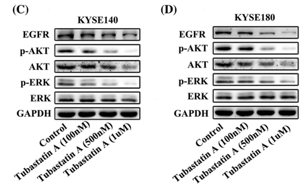
|
29665050 | |
| Immunofluorescence | α-tubulin / Acetylated tubulin HDAC6 |
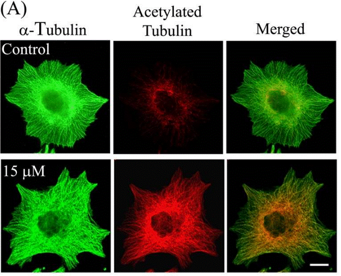
|
23798680 | |
| In Vivo | ||
| In Vivo | Tubastatin A reduces the growth of cholangiocarcinoma in vivo. This compound (10 mg/kg) induces a 6-fold lower mean tumor weights in syngeneic rat orthotopic model of cholangiocarcinoma, and reduction of the ratios of tumor weight to liver weight and body weight (5- and 5.6-fold, respectively), as well as a greater frequency of ciliated cholangiocytes compared with controls (29% vs 1.4%). It significantly decreases the amount of PCNA-positive cells in the treated tumors compared with vehicle controls (34% vs 65%). This chemical shows significant inhibition of paw volume at 30 mg/kg i.p. in a Freund's complete adjuvant (FCA) induced animal model of inflammation. It (30 mg/kg i.p.) significant attenuates clinical scores (~ 70%), and IL-6 expression in paw tissues of collagen induced arthritis DBA1 mouse. | |
|---|---|---|
| 動物実験 | 動物モデル | Rat cholangiocarcinoma xenografts BDEneu |
| 投与量 | 10 mg/kg | |
| 投与経路 | i.p. daily | |
|
化学情報
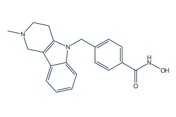
| 分子量 | 335.4 | 化学式 | C20H21N3O2 |
| CAS No. | 1252003-15-8 | SDF | Download Tubastatin A SDFをダウンロードする |
| Smiles | CN1CCC2=C(C1)C3=CC=CC=C3N2CC4=CC=C(C=C4)C(=O)NO | ||
| 保管 | |||
|
In vitro |
DMSO : 67 mg/mL ( (199.76 mM); 吸湿したDMSOは溶解度を減少させます。新しいDMSOをご使用ください。) Water : Insoluble Ethanol : Insoluble |
モル濃度計算器 |
|
in vivo Add solvents to the product individually and in order. |
投与溶液組成計算機 | |||||
実験計算
投与溶液組成計算機(クリア溶液)
ステップ1:実験データを入力してください。(実験操作によるロスを考慮し、動物数を1匹分多くして計算・調製することを推奨します)
mg/kg
g
μL
匹
ステップ2:投与溶媒の組成を入力してください。(ロット毎に適した溶解組成が異なる場合があります。詳細については弊社までお問い合わせください)
% DMSO
%
% Tween 80
% ddH2O
%DMSO
%
計算結果:
投与溶媒濃度: mg/ml;
DMSOストック溶液調製方法: mg 試薬を μL DMSOに溶解する(濃度 mg/mL, 注:濃度が当該ロットのDMSO溶解度を超える場合はご連絡ください。 )
投与溶媒調製方法:Take μL DMSOストック溶液に μL PEG300,を加え、完全溶解後μL Tween 80,を加えて完全溶解させた後 μL ddH2O,を加え完全に溶解させます。
投与溶媒調製方法:μL DMSOストック溶液に μL Corn oil,を加え、完全溶解。
注意:1.ストック溶液に沈殿、混濁などがないことをご確認ください;
2.順番通りに溶剤を加えてください。次のステップに進む前に溶液に沈殿、混濁などがないことを確認してから加えてください。ボルテックス、ソニケーション、水浴加熱など物理的な方法で溶解を早めることは可能です。
技術サポート
ストックの作り方、阻害剤の保管方法、細胞実験や動物実験の際に注意すべき点など、製品を取扱う時に問い合わせが多かった質問に対しては取扱説明書でお答えしています。
他に質問がある場合は、お気軽にお問い合わせください。
* 必須
よくある質問(FAQ)
質問1:
We are planning to order some, but I found out there are two versions of it. One has HCl and one does not. Which one do you recommend for live cell use? Will the HCl containing version significantly change the pH?
回答
S8049 and S2627 have the same molecular structure. The only difference is S2627 containing HCl and has higher solubility in DMSO (74 mg/mL vs. S8049 9 mg/mL). Since they are the same molecule, its biological function should be similar. I would recommend to use S2627 for cell culture study.
質問2:
What vehicle do you recommend to dissolve it for in vivo experiments?
回答
It can be dissolved in 2% DMSO/30% PEG 300/PBS at 2.5 mg/mL as a clear solution, and is also a clear solution in 2% DMSO/ corn oil at 2.5 mg/mL. This compound in 2% DMSO/0.5% Tween 80/PBS is a homogeneous suspension at 2.5 mg/mL at first. After stay for a while, the precipitation goes out at the bottom of the tube.
納期 国内在庫品:受注日の翌日(15時までの受注分) *北海道、九州、沖縄への配送は受注日より2日以上 を要する場合あり 海外在庫品:受注後1〜2週間